 Movie
Movie Controller

[English] 日本語
 Yorodumi
- PDB-4b5u: Crystal structures of divalent metal dependent pyruvate aldolase,... -
+ Open data

- Basic information
| Entry | Database: PDB / ID: 4b5u | |||||||||
|---|---|---|---|---|---|---|---|---|---|---|
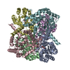
| Title | Crystal structures of divalent metal dependent pyruvate aldolase, HpaI, in complex with pyruvate and succinic semialdehyde | |||||||||
 Components Components | 4-HYDROXY-2-OXO-HEPTANE-1,7-DIOATE ALDOLASE | |||||||||
 Keywords Keywords | LYASE / CATALYTIC MECHANISM | |||||||||
| Function / homology |  Function and homology information Function and homology information4-hydroxy-2-ketopimelate aldolase activity / 4-hydroxyphenylacetate catabolic process / 4-hydroxy-2-oxoheptanedioate aldolase / 2-dehydro-3-deoxy-D-gluconate aldolase activity / phenylacetate catabolic process / metal ion binding / cytoplasm Similarity search - Function | |||||||||
| Biological species |  | |||||||||
| Method |  X-RAY DIFFRACTION / SYNCHROTRON / MOLECULAR REPLACEMENT / Resolution: 1.913 Å X-RAY DIFFRACTION / SYNCHROTRON / MOLECULAR REPLACEMENT / Resolution: 1.913 Å | |||||||||
 Authors Authors | Coincon, M. / Wang, W. / Seah, S.Y.K. / Sygusch, J. | |||||||||
 Citation Citation | Journal: J.Biol.Chem. / Year: 2012 Title: Crystal Structure of Reaction Intermediates in Pyruvate Class II Aldolase: Substrate Cleavage, Enolate Stabilization and Substrate Specificity Authors: Coincon, M. / Wang, W. / Sygusch, J. / Seah, S.Y.K. | |||||||||
| History |
| |||||||||
| Remark 700 | SHEET DETERMINATION METHOD: DSSP THE SHEETS PRESENTED AS "AA" IN EACH CHAIN ON SHEET RECORDS BELOW ... SHEET DETERMINATION METHOD: DSSP THE SHEETS PRESENTED AS "AA" IN EACH CHAIN ON SHEET RECORDS BELOW IS ACTUALLY AN 8-STRANDED BARREL THIS IS REPRESENTED BY A 9-STRANDED SHEET IN WHICH THE FIRST AND LAST STRANDS ARE IDENTICAL. THE SHEETS PRESENTED AS "BA" IN EACH CHAIN ON SHEET RECORDS BELOW IS ACTUALLY AN 8-STRANDED BARREL THIS IS REPRESENTED BY A 9-STRANDED SHEET IN WHICH THE FIRST AND LAST STRANDS ARE IDENTICAL. |
- Structure visualization
| Structure viewer | Molecule: MolmilJmol/JSmol |
|---|
- Downloads & links
-Download
| PDBx/mmCIF format | 4b5u.cif.gz | 212.1 KB | Display | PDBx/mmCIF format |
|---|---|---|---|---|
| PDB format | pdb4b5u.ent.gz | 171.1 KB | Display | PDB format |
| PDBx/mmJSON format | 4b5u.json.gz | Tree view | PDBx/mmJSON format | |
| Others |  Other downloads Other downloads |
-Validation report
| Arichive directory | https://data.pdbj.org/pub/pdb/validation_reports/b5/4b5uftp://data.pdbj.org/pub/pdb/validation_reports/b5/4b5u | HTTPS FTP |
|---|
-Related structure data
| Related structure data |  4b5sC 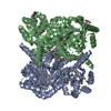 4b5tC 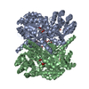 4b5vC 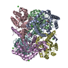 4b5wC 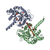 4b5xC  2v5jS C: citing same article ( S: Starting model for refinement |
|---|---|
| Similar structure data |


-Links
 PDBj
PDBj


- Assembly
| Deposited unit | 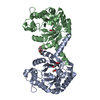
| ||||||||||||
|---|---|---|---|---|---|---|---|---|---|---|---|---|---|
| 1 | 
| ||||||||||||
| Unit cell |
| ||||||||||||
| Components on special symmetry positions |
|
-Components
-Protein , 1 types, 2 molecules AB
| #1: Protein | Mass: 27009.793 Da / Num. of mol.: 2 / Fragment: RESIDUES 1-251 Source method: isolated from a genetically manipulated source Source: (gene. exp.) References: UniProt: B1IS70, 2-dehydro-3-deoxyglucarate aldolase |
|---|
-Non-polymers , 6 types, 818 molecules 


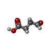

| #2: Chemical |  Mass: 58.933 Da / Num. of mol.: 2 / Source method: obtained synthetically / Formula: Co Mass: 58.933 Da / Num. of mol.: 2 / Source method: obtained synthetically / Formula: Co#3: Chemical |  Mass: 24.305 Da / Num. of mol.: 2 / Source method: obtained synthetically / Formula: Mg Mass: 24.305 Da / Num. of mol.: 2 / Source method: obtained synthetically / Formula: Mg#4: Chemical |  Mass: 92.094 Da / Num. of mol.: 2 / Source method: obtained synthetically / Formula: C3H8O3 Mass: 92.094 Da / Num. of mol.: 2 / Source method: obtained synthetically / Formula: C3H8O3#5: Chemical |  Mass: 88.062 Da / Num. of mol.: 2 / Source method: obtained synthetically / Formula: C3H4O3 Mass: 88.062 Da / Num. of mol.: 2 / Source method: obtained synthetically / Formula: C3H4O3#6: Chemical |  Mass: 102.089 Da / Num. of mol.: 2 / Source method: obtained synthetically / Formula: C4H6O3 Mass: 102.089 Da / Num. of mol.: 2 / Source method: obtained synthetically / Formula: C4H6O3#7: Water | ChemComp-HOH / | Mass: 18.015 Da / Num. of mol.: 808 / Source method: isolated from a natural source / Formula: H2O |
|---|
-Experimental details
-Experiment
| Experiment | Method: X-RAY DIFFRACTION / Number of used crystals: 1 |
|---|
- Sample preparation
| Crystal | Density Matthews: 4.02 Å3/Da / Density % sol: 69.17 % / Description: WE REPORTED RPIM INSTEAD OF RMERGE |
|---|---|
| Crystal grow | Details: 0.5 M AMMONIUM DIHYDROGEN PHOSPHATE, 30% GLYCEROL, 2 MM COCL2 PH 5 |
-Data collection
| Diffraction | Mean temperature: 100 K |
|---|---|
| Diffraction source | Source: SYNCHROTRON / Site: NSLS  / Beamline: X25 / Wavelength: 0.9795 / Beamline: X25 / Wavelength: 0.9795 |
| Detector | Type: ADSC QUANTUM 315 / Detector: CCD |
| Radiation | Protocol: SINGLE WAVELENGTH / Monochromatic (M) / Laue (L): M / Scattering type: x-ray |
| Radiation wavelength | Wavelength: 0.9795 Å / Relative weight: 1 |
| Reflection | Resolution: 1.91→41.9 Å / Num. obs: 87289 / % possible obs: 98.6 % / Observed criterion σ(I): 1 / Redundancy: 21.8 % / Biso Wilson estimate: 18.75 Å2 / Rmerge(I) obs: 0.03 / Net I/σ(I): 21.1 |
| Reflection shell | Resolution: 1.91→1.96 Å / Redundancy: 4.9 % / Rmerge(I) obs: 0.24 / Mean I/σ(I) obs: 2.9 / % possible all: 81.7 |
- Processing
| Software |
| |||||||||||||||||||||||||||||||||||||||||||||||||||||||||||||||||||||||||||||||||||||||||||||||||||||||||||||||||||||||||||||||||||||||||||||||||||||||||||||||||||||||||||||||||||||||||||||||||||||||||||||||||||||||||
|---|---|---|---|---|---|---|---|---|---|---|---|---|---|---|---|---|---|---|---|---|---|---|---|---|---|---|---|---|---|---|---|---|---|---|---|---|---|---|---|---|---|---|---|---|---|---|---|---|---|---|---|---|---|---|---|---|---|---|---|---|---|---|---|---|---|---|---|---|---|---|---|---|---|---|---|---|---|---|---|---|---|---|---|---|---|---|---|---|---|---|---|---|---|---|---|---|---|---|---|---|---|---|---|---|---|---|---|---|---|---|---|---|---|---|---|---|---|---|---|---|---|---|---|---|---|---|---|---|---|---|---|---|---|---|---|---|---|---|---|---|---|---|---|---|---|---|---|---|---|---|---|---|---|---|---|---|---|---|---|---|---|---|---|---|---|---|---|---|---|---|---|---|---|---|---|---|---|---|---|---|---|---|---|---|---|---|---|---|---|---|---|---|---|---|---|---|---|---|---|---|---|---|---|---|---|---|---|---|---|---|---|---|---|---|---|---|---|---|
| Refinement | Method to determine structure: MOLECULAR REPLACEMENT Starting model: PDB ENTRY 2V5J Resolution: 1.913→41.916 Å / SU ML: 0.13 / σ(F): 2 / Phase error: 12.9 / Stereochemistry target values: ML
| |||||||||||||||||||||||||||||||||||||||||||||||||||||||||||||||||||||||||||||||||||||||||||||||||||||||||||||||||||||||||||||||||||||||||||||||||||||||||||||||||||||||||||||||||||||||||||||||||||||||||||||||||||||||||
| Solvent computation | Shrinkage radii: 1.4 Å / VDW probe radii: 1.3 Å / Solvent model: FLAT BULK SOLVENT MODEL / Bsol: 4 Å2 / ksol: 4 e/Å3 | |||||||||||||||||||||||||||||||||||||||||||||||||||||||||||||||||||||||||||||||||||||||||||||||||||||||||||||||||||||||||||||||||||||||||||||||||||||||||||||||||||||||||||||||||||||||||||||||||||||||||||||||||||||||||
| Displacement parameters | Biso mean: 23.9 Å2 | |||||||||||||||||||||||||||||||||||||||||||||||||||||||||||||||||||||||||||||||||||||||||||||||||||||||||||||||||||||||||||||||||||||||||||||||||||||||||||||||||||||||||||||||||||||||||||||||||||||||||||||||||||||||||
| Refinement step | Cycle: LAST / Resolution: 1.913→41.916 Å
| |||||||||||||||||||||||||||||||||||||||||||||||||||||||||||||||||||||||||||||||||||||||||||||||||||||||||||||||||||||||||||||||||||||||||||||||||||||||||||||||||||||||||||||||||||||||||||||||||||||||||||||||||||||||||
| Refine LS restraints |
| |||||||||||||||||||||||||||||||||||||||||||||||||||||||||||||||||||||||||||||||||||||||||||||||||||||||||||||||||||||||||||||||||||||||||||||||||||||||||||||||||||||||||||||||||||||||||||||||||||||||||||||||||||||||||
| LS refinement shell |
|
