 Movie
Movie Controller

[English] 日本語
 Yorodumi
- PDB-1ybo: Crystal structure of the PDZ tandem of human syntenin with syndec... -
+ Open data

- Basic information
| Entry | Database: PDB / ID: 1ybo | ||||||
|---|---|---|---|---|---|---|---|
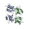
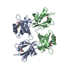
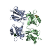
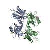
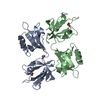
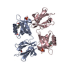
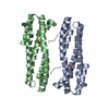
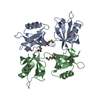
| Title | Crystal structure of the PDZ tandem of human syntenin with syndecan peptide | ||||||
 Components Components |
| ||||||
 Keywords Keywords | STRUCTURAL PROTEIN / PDZ domain / scaffolding protein / adhesion complex | ||||||
| Function / homology |  Function and homology information Function and homology informationDefective B3GALT6 causes EDSP2 and SEMDJL1 / Defective B4GALT7 causes EDS, progeroid type / Defective B3GAT3 causes JDSSDHD / Defective EXT2 causes exostoses 2 / Defective EXT1 causes exostoses 1, TRPS2 and CHDS / regulation of fibroblast migration / Glycosaminoglycan-protein linkage region biosynthesis / interleukin-5 receptor complex / HS-GAG biosynthesis / interleukin-5 receptor binding ...Defective B3GALT6 causes EDSP2 and SEMDJL1 / Defective B4GALT7 causes EDS, progeroid type / Defective B3GAT3 causes JDSSDHD / Defective EXT2 causes exostoses 2 / Defective EXT1 causes exostoses 1, TRPS2 and CHDS / regulation of fibroblast migration / Glycosaminoglycan-protein linkage region biosynthesis / interleukin-5 receptor complex / HS-GAG biosynthesis / interleukin-5 receptor binding / positive regulation of extracellular exosome assembly / inner ear receptor cell stereocilium organization / Neurofascin interactions / HS-GAG degradation / syndecan binding / cytoskeletal anchor activity / substrate-dependent cell migration, cell extension / positive regulation of exosomal secretion / costamere / negative regulation of receptor internalization / frizzled binding / Ephrin signaling / ureteric bud development / protein targeting to membrane / RIPK1-mediated regulated necrosis / Syndecan interactions / thrombospondin receptor activity / positive regulation of transforming growth factor beta receptor signaling pathway / RSV-host interactions / fibronectin binding / Respiratory syncytial virus (RSV) attachment and entry / positive regulation of phosphorylation / positive regulation of focal adhesion assembly / positive regulation of epithelial to mesenchymal transition / Retinoid metabolism and transport / negative regulation of T cell proliferation / positive regulation of stress fiber assembly / phosphatidylinositol-4,5-bisphosphate binding / protein sequestering activity / lysosomal lumen / regulation of mitotic cell cycle / protein kinase C binding / Cell surface interactions at the vascular wall / adherens junction / neural tube closure / wound healing / positive regulation of JNK cascade / Golgi lumen / Regulation of necroptotic cell death / azurophil granule lumen / melanosome / cell migration / extracellular vesicle / positive regulation of cell growth / actin cytoskeleton organization / blood microparticle / nuclear membrane / chemical synaptic transmission / Ras protein signal transduction / Attachment and Entry / cytoskeleton / intracellular signal transduction / positive regulation of cell migration / membrane raft / protein heterodimerization activity / focal adhesion / positive regulation of cell population proliferation / Neutrophil degranulation / synapse / endoplasmic reticulum membrane / cell surface / negative regulation of transcription by RNA polymerase II / extracellular space / extracellular exosome / extracellular region / nucleoplasm / identical protein binding / membrane / nucleus / plasma membrane / cytoplasm / cytosol Similarity search - Function | ||||||
| Biological species |  Homo sapiens (human) Homo sapiens (human) | ||||||
| Method |  X-RAY DIFFRACTION / SYNCHROTRON / MOLECULAR REPLACEMENT / Resolution: 2.3 Å X-RAY DIFFRACTION / SYNCHROTRON / MOLECULAR REPLACEMENT / Resolution: 2.3 Å | ||||||
 Authors Authors | Grembecka, J. / Cooper, D.R. / Cierpicki, T. / Kang, B.S. / Devedjiev, Y. / Derewenda, Z. | ||||||
 Citation Citation | Journal: Biochemistry / Year: 2006 Title: The binding of the PDZ tandem of syntenin to target proteins Authors: Grembecka, J. / Cierpicki, T. / Devedjiev, Y. / Derewenda, U. / Kang, B.S. / Bushweller, J.H. / Derewenda, Z.S. | ||||||
| History |
|
- Structure visualization
| Structure viewer | Molecule: MolmilJmol/JSmol |
|---|
- Downloads & links
-Download
| PDBx/mmCIF format | 1ybo.cif.gz | 81.1 KB | Display | PDBx/mmCIF format |
|---|---|---|---|---|
| PDB format | pdb1ybo.ent.gz | 61.1 KB | Display | PDB format |
| PDBx/mmJSON format | 1ybo.json.gz | Tree view | PDBx/mmJSON format | |
| Others |  Other downloads Other downloads |
-Validation report
| Arichive directory | https://data.pdbj.org/pub/pdb/validation_reports/yb/1yboftp://data.pdbj.org/pub/pdb/validation_reports/yb/1ybo | HTTPS FTP |
|---|
-Related structure data
| Related structure data |  1v1tC 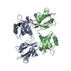 1w9eC 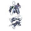 1w9oC 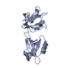 1w9qC  1n99S S: Starting model for refinement C: citing same article ( |
|---|---|
| Similar structure data |


-Links
 PDBj
PDBj


- Assembly
| Deposited unit | 
| ||||||||
|---|---|---|---|---|---|---|---|---|---|
| 1 |
| ||||||||
| Unit cell |
| ||||||||
| Details | The dimer in the assymetric unit is the biological assembly |
-Components
| #1: Protein | Mass: 18018.688 Da / Num. of mol.: 2 Source method: isolated from a genetically manipulated source Source: (gene. exp.) Homo sapiens (human) / Gene: SDCBP, MDA9, SYCL / Plasmid: pGST-parallel-1 / Species (production host): Escherichia coli / Production host:  #2: Protein/peptide | Mass: 1972.329 Da / Num. of mol.: 2 / Source method: obtained synthetically Details: synthesized peptide cooresponding to the C-terminus of syndecan References: UniProt: P31431 #3: Water | ChemComp-HOH / |  Mass: 18.015 Da / Num. of mol.: 190 / Source method: isolated from a natural source / Formula: H2O Mass: 18.015 Da / Num. of mol.: 190 / Source method: isolated from a natural source / Formula: H2O |
|---|
-Experimental details
-Experiment
| Experiment | Method: X-RAY DIFFRACTION / Number of used crystals: 1 |
|---|
- Sample preparation
| Crystal | Density Matthews: 2.3 Å3/Da / Density % sol: 49.7 % |
|---|---|
| Crystal grow | Method: vapor diffusion, sitting drop Details: 20% PEG 3350, 0.2M NH4Cl, VAPOR DIFFUSION, SITTING DROP |
-Data collection
| Diffraction | Mean temperature: 100 K |
|---|---|
| Diffraction source | Source: SYNCHROTRON / Site: NSLS  / Beamline: X8C / Wavelength: 0.9796 Å / Beamline: X8C / Wavelength: 0.9796 Å |
| Detector | Type: ADSC QUANTUM 4 / Detector: CCD / Date: Apr 11, 2003 |
| Radiation | Protocol: SINGLE WAVELENGTH / Monochromatic (M) / Laue (L): M / Scattering type: x-ray |
| Radiation wavelength | Wavelength: 0.9796 Å / Relative weight: 1 |
| Reflection | Resolution: 2→40 Å / Num. obs: 23083 / % possible obs: 99 % / Redundancy: 8.3 % / Biso Wilson estimate: 25.7 Å2 / Rmerge(I) obs: 0.07 / Χ2: 1.388 / Net I/σ(I): 30.3 |
| Reflection shell | Resolution: 2→2.07 Å / Redundancy: 7.9 % / Rmerge(I) obs: 0.283 / Mean I/σ(I) obs: 8.31 / Num. unique all: 2282 / Χ2: 1.208 / % possible all: 100 |
- Processing
| Software |
| ||||||||||||||||||||||||||||||||||||||||||||||||||||||||||||||||||||||||||||||||||||||||||||||||||||||||||||||||||||||||||||||||||
|---|---|---|---|---|---|---|---|---|---|---|---|---|---|---|---|---|---|---|---|---|---|---|---|---|---|---|---|---|---|---|---|---|---|---|---|---|---|---|---|---|---|---|---|---|---|---|---|---|---|---|---|---|---|---|---|---|---|---|---|---|---|---|---|---|---|---|---|---|---|---|---|---|---|---|---|---|---|---|---|---|---|---|---|---|---|---|---|---|---|---|---|---|---|---|---|---|---|---|---|---|---|---|---|---|---|---|---|---|---|---|---|---|---|---|---|---|---|---|---|---|---|---|---|---|---|---|---|---|---|---|---|
| Refinement | Method to determine structure: MOLECULAR REPLACEMENT Starting model: 1N99 Resolution: 2.3→40 Å / Cor.coef. Fo:Fc: 0.93 / Cor.coef. Fo:Fc free: 0.868 / SU B: 14.26 / SU ML: 0.189 / SU R Cruickshank DPI: 0.403 / TLS residual ADP flag: LIKELY RESIDUAL / Cross valid method: THROUGHOUT / ESU R Free: 0.28 / Stereochemistry target values: Engh & Huber
| ||||||||||||||||||||||||||||||||||||||||||||||||||||||||||||||||||||||||||||||||||||||||||||||||||||||||||||||||||||||||||||||||||
| Solvent computation | Ion probe radii: 0.8 Å / Shrinkage radii: 0.8 Å / VDW probe radii: 1.2 Å / Solvent model: MASK | ||||||||||||||||||||||||||||||||||||||||||||||||||||||||||||||||||||||||||||||||||||||||||||||||||||||||||||||||||||||||||||||||||
| Displacement parameters | Biso mean: 25.247 Å2
| ||||||||||||||||||||||||||||||||||||||||||||||||||||||||||||||||||||||||||||||||||||||||||||||||||||||||||||||||||||||||||||||||||
| Refinement step | Cycle: LAST / Resolution: 2.3→40 Å
| ||||||||||||||||||||||||||||||||||||||||||||||||||||||||||||||||||||||||||||||||||||||||||||||||||||||||||||||||||||||||||||||||||
| Refine LS restraints |
| ||||||||||||||||||||||||||||||||||||||||||||||||||||||||||||||||||||||||||||||||||||||||||||||||||||||||||||||||||||||||||||||||||
| LS refinement shell | Resolution: 2.3→2.36 Å / Total num. of bins used: 20
| ||||||||||||||||||||||||||||||||||||||||||||||||||||||||||||||||||||||||||||||||||||||||||||||||||||||||||||||||||||||||||||||||||
| Refinement TLS params. | Method: refined / Refine-ID: X-RAY DIFFRACTION
| ||||||||||||||||||||||||||||||||||||||||||||||||||||||||||||||||||||||||||||||||||||||||||||||||||||||||||||||||||||||||||||||||||
| Refinement TLS group | Refine-ID: X-RAY DIFFRACTION / Selection: ALL
|
