 Movie
Movie Controller

[English] 日本語
 Yorodumi
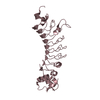
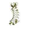
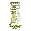
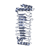
- PDB-1y8x: Structural basis for recruitment of Ubc12 by an E2-binding domain... -
+ Open data

- Basic information
| Entry | Database: PDB / ID: 1y8x | ||||||
|---|---|---|---|---|---|---|---|
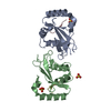
| Title | Structural basis for recruitment of Ubc12 by an E2-binding domain in NEDD8's E1 | ||||||
 Components Components |
| ||||||
 Keywords Keywords |  LIGASE / Ubiquitin-conjugating enzyme E2 M LIGASE / Ubiquitin-conjugating enzyme E2 M | ||||||
| Function / homology |  Function and homology information Function and homology informationE2 NEDD8-conjugating enzyme / NEDD8 conjugating enzyme activity / E1 NEDD8-activating enzyme / NEDD8 activating enzyme activity / endomitotic cell cycle / NEDD8 transferase activity / protein neddylation / TGF-beta receptor signaling activates SMADs / post-translational protein modification / NIK-->noncanonical NF-kB signaling ...E2 NEDD8-conjugating enzyme / NEDD8 conjugating enzyme activity / E1 NEDD8-activating enzyme / NEDD8 activating enzyme activity / endomitotic cell cycle / NEDD8 transferase activity / protein neddylation / TGF-beta receptor signaling activates SMADs / post-translational protein modification / NIK-->noncanonical NF-kB signaling / Dectin-1 mediated noncanonical NF-kB signaling / protein modification process / ubiquitin-protein transferase activity / positive regulation of neuron apoptotic process / Antigen processing: Ubiquitination & Proteasome degradation / Neddylation / regulation of cell cycle / protein heterodimerization activity / protein-containing complex / proteolysis / nucleoplasm / ATP binding / identical protein binding / nucleus / cytosol / cytoplasmSimilarity search - Function | ||||||
| Biological species |  Homo sapiens (human) Homo sapiens (human) | ||||||
| Method | X-RAY DIFFRACTION / SYNCHROTRON / MAD / Resolution: 2.4 Å | ||||||
 Authors Authors | Huang, D.T. / Paydar, A. / Zhuang, M. / Waddell, M.B. / Holton, J.M. / Schulman, B.A. | ||||||
 Citation Citation | Journal: Mol.Cell / Year: 2005 Title: Structural basis for recruitment of Ubc12 by an E2 binding domain in NEDD8's E1. Authors: Huang, D.T. / Paydar, A. / Zhuang, M. / Waddell, M.B. / Holton, J.M. / Schulman, B.A. | ||||||
| History |
|
- Structure visualization
| Structure viewer | Molecule: MolmilJmol/JSmol |
|---|
- Downloads & links
-Download
| PDBx/mmCIF format | 1y8x.cif.gz | 61.2 KB | Display | PDBx/mmCIF format |
|---|---|---|---|---|
| PDB format | pdb1y8x.ent.gz | 48.1 KB | Display | PDB format |
| PDBx/mmJSON format | 1y8x.json.gz | Tree view | PDBx/mmJSON format | |
| Others |  Other downloads Other downloads |
-Validation report
| Arichive directory | https://data.pdbj.org/pub/pdb/validation_reports/y8/1y8xftp://data.pdbj.org/pub/pdb/validation_reports/y8/1y8x | HTTPS FTP |
|---|
-Related structure data
| Similar structure data |
|---|


-Links
 PDBj
PDBj


- Assembly
| Deposited unit | 
| ||||||||||
|---|---|---|---|---|---|---|---|---|---|---|---|
| 1 |
| ||||||||||
| Unit cell |
|
-Components
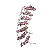
| #1: Protein | Mass: 18485.615 Da / Num. of mol.: 1 Source method: isolated from a genetically manipulated source Source: (gene. exp.) Homo sapiens (human) / Gene: UBE2M, UBC12 / Production host:  Escherichia coli (E. coli) / References: UniProt: P61081, ubiquitin-protein ligase Escherichia coli (E. coli) / References: UniProt: P61081, ubiquitin-protein ligase |
|---|---|
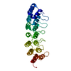
| #2: Protein | Mass: 10856.974 Da / Num. of mol.: 1 Source method: isolated from a genetically manipulated source Source: (gene. exp.) Homo sapiens (human) / Gene: UBE1C, UBA3 / Production host: Escherichia coli (E. coli) / References: UniProt: Q8TBC4 |
| #3: Water | ChemComp-HOH / Water Mass: 18.015 Da / Num. of mol.: 112 / Source method: isolated from a natural source / Formula: H2O Mass: 18.015 Da / Num. of mol.: 112 / Source method: isolated from a natural source / Formula: H2O |
-Experimental details
-Experiment
| Experiment | Method: X-RAY DIFFRACTION |
|---|
- Sample preparation
| Crystal | Density Matthews: 2.68 Å3/Da / Density % sol: 54.11 % |
|---|
-Data collection
| Diffraction |
| ||||||||||||||||||||||||||||||
|---|---|---|---|---|---|---|---|---|---|---|---|---|---|---|---|---|---|---|---|---|---|---|---|---|---|---|---|---|---|---|---|
| Diffraction source |
| ||||||||||||||||||||||||||||||
| Radiation | Protocol: MAD / Monochromatic (M) / Laue (L): M / Scattering type: x-ray | ||||||||||||||||||||||||||||||
| Radiation wavelength |
| ||||||||||||||||||||||||||||||
| Reflection | Resolution: 2.4→50 Å / Num. all: 12655 / Num. obs: 12655 / Redundancy: 11.4 % / Rsym value: 0.099 / Net I/σ(I): 38.3 |

- Processing
| Refinement | Method to determine structure: MAD / Resolution: 2.4→50 Å / σ(F): 0 / Stereochemistry target values: Engh & Huber
| ||||||||||||||||||||
|---|---|---|---|---|---|---|---|---|---|---|---|---|---|---|---|---|---|---|---|---|---|
| Refinement step | Cycle: LAST / Resolution: 2.4→50 Å
|
