 Movie
Movie Controller

[English] 日本語
 Yorodumi
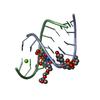
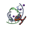
- PDB-1y84: Crystal structure of the A-DNA GCGTAT*CGC with a 2'-O-[2-(imidazo... -
+ Open data

- Basic information
| Entry | Database: PDB / ID: 1y84 | ||||||||||||||||||
|---|---|---|---|---|---|---|---|---|---|---|---|---|---|---|---|---|---|---|---|
| Title | Crystal structure of the A-DNA GCGTAT*CGC with a 2'-O-[2-(imidazolyl)ethyl] Thymidine (T*) | ||||||||||||||||||
 Components Components | 5'-D(* Keywords KeywordsDNA / A-DNA / O2'-modification / decamer. | Function / homology | SPERMINE / DNA |  Function and homology information Function and homology informationMethod |  X-RAY DIFFRACTION / MOLECULAR REPLACEMENT / Resolution: 1.6 Å X-RAY DIFFRACTION / MOLECULAR REPLACEMENT / Resolution: 1.6 Å  Authors AuthorsEgli, M. / Minasov, G. / Tereshko, V. / Pallan, P.S. / Teplova, M. / Inamati, G.B. / Lesnik, E.A. / Owens, S.R. / Ross, B.S. / Prakash, T.P. / Manoharan, M. |  CitationJournal: Biochemistry / Year: 2005 CitationJournal: Biochemistry / Year: 2005Title: Probing the Influence of Stereoelectronic Effects on the Biophysical Properties of Oligonucleotides: Comprehensive Analysis of the RNA Affinity, Nuclease Resistance, and Crystal Structure of ...Title: Probing the Influence of Stereoelectronic Effects on the Biophysical Properties of Oligonucleotides: Comprehensive Analysis of the RNA Affinity, Nuclease Resistance, and Crystal Structure of Ten 2'-O-Ribonucleic Acid Modifications. Authors: Egli, M. / Minasov, G. / Tereshko, V. / Pallan, P.S. / Teplova, M. / Inamati, G.B. / Lesnik, E.A. / Owens, S.R. / Ross, B.S. / Prakash, T.P. / Manoharan, M. History |
|
- Structure visualization
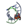
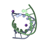
| Structure viewer | Molecule: MolmilJmol/JSmol |
|---|
- Downloads & links
-Download
| PDBx/mmCIF format | 1y84.cif.gz | 26.1 KB | Display | PDBx/mmCIF format |
|---|---|---|---|---|
| PDB format | pdb1y84.ent.gz | 16.5 KB | Display | PDB format |
| PDBx/mmJSON format | 1y84.json.gz | Tree view | PDBx/mmJSON format | |
| Others |  Other downloads Other downloads |
-Validation report
| Arichive directory | https://data.pdbj.org/pub/pdb/validation_reports/y8/1y84ftp://data.pdbj.org/pub/pdb/validation_reports/y8/1y84 | HTTPS FTP |
|---|
-Related structure data
| Related structure data | 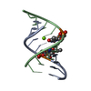 1wv5C 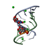 1wv6C  1y7fC  1y86C  1y8lC 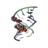 1y8vC  1y9fC 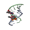 1y9sC 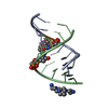 1yb9C 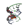 1ybcC  410dS C: citing same article ( S: Starting model for refinement |
|---|---|
| Similar structure data |


-Links
 PDBj
PDBj


- Assembly
| Deposited unit | 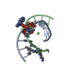
| ||||||||
|---|---|---|---|---|---|---|---|---|---|
| 1 |
| ||||||||
| Unit cell |
| ||||||||
| Details | Chains A and B form duplex |
-Components
| #1: DNA chain | Mass: 3155.119 Da / Num. of mol.: 2 / Source method: obtained synthetically #2: Chemical | ChemComp-MG / |   Mass: 24.305 Da / Num. of mol.: 1 / Source method: obtained synthetically / Formula: Mg Mass: 24.305 Da / Num. of mol.: 1 / Source method: obtained synthetically / Formula: Mg#3: Chemical | ChemComp-SPM / |   Mass: 202.340 Da / Num. of mol.: 1 / Source method: obtained synthetically / Formula: C10H26N4 Mass: 202.340 Da / Num. of mol.: 1 / Source method: obtained synthetically / Formula: C10H26N4#4: Water | ChemComp-HOH / |  Mass: 18.015 Da / Num. of mol.: 114 / Source method: isolated from a natural source / Formula: H2O Mass: 18.015 Da / Num. of mol.: 114 / Source method: isolated from a natural source / Formula: H2O |
|---|
-Experimental details
-Experiment
| Experiment | Method: X-RAY DIFFRACTION / Number of used crystals: 1 |
|---|
- Sample preparation
| Crystal | Density Matthews: 1.9 Å3/Da / Density % sol: 35.34 % | ||||||||||||||||||||||||||||||||||||||||||||||||||||
|---|---|---|---|---|---|---|---|---|---|---|---|---|---|---|---|---|---|---|---|---|---|---|---|---|---|---|---|---|---|---|---|---|---|---|---|---|---|---|---|---|---|---|---|---|---|---|---|---|---|---|---|---|---|
| Crystal grow | Temperature: 295 K / Method: vapor diffusion, hanging drop / pH: 6 Details: 10%MPD, 40mM Na-Cacodilate, 12 mM Spermine, 80mM NaCL, 12mM KCL, 20mM MgCL2, pH 6.0, VAPOR DIFFUSION, HANGING DROP, temperature 295K | ||||||||||||||||||||||||||||||||||||||||||||||||||||
| Components of the solutions |
|
-Data collection
| Diffraction | Mean temperature: 110 K |
|---|---|
| Diffraction source | Source: ROTATING ANODE / Type: RIGAKU RU200 / Wavelength: 1.5418 |
| Detector | Type: RIGAKU RAXIS IIC / Detector: IMAGE PLATE / Date: Jul 6, 1999 / Details: Mirrors |
| Radiation | Protocol: SINGLE WAVELENGTH / Monochromatic (M) / Laue (L): M / Scattering type: x-ray |
| Radiation wavelength | Wavelength: 1.5418 Å / Relative weight: 1 |
| Reflection | Resolution: 1.6→25 Å / Num. all: 6710 / Num. obs: 6710 / % possible obs: 99.2 % / Observed criterion σ(I): -3 / Redundancy: 4.2 % / Rmerge(I) obs: 0.096 / Net I/σ(I): 12.8 |
| Reflection shell | Resolution: 1.6→1.66 Å / Redundancy: 2.3 % / Rmerge(I) obs: 0.376 / Mean I/σ(I) obs: 2.9 / Num. unique all: 619 / % possible all: 95.5 |
- Processing
| Software |
| |||||||||||||||||||||||||
|---|---|---|---|---|---|---|---|---|---|---|---|---|---|---|---|---|---|---|---|---|---|---|---|---|---|---|
| Refinement | Method to determine structure: MOLECULAR REPLACEMENT Starting model: PDB entry 410D Resolution: 1.6→20 Å / Isotropic thermal model: Isotropic / Cross valid method: THROUGHOUT / σ(F): 0 / σ(I): 0 Details: Conjugate gradient refinement using maximum likelihood target for amplitudes
| |||||||||||||||||||||||||
| Displacement parameters | Biso mean: 21.3 Å2
| |||||||||||||||||||||||||
| Refinement step | Cycle: LAST / Resolution: 1.6→20 Å
| |||||||||||||||||||||||||
| Refine LS restraints |
| |||||||||||||||||||||||||
| LS refinement shell | Resolution: 1.6→1.64 Å
|
