| 登録情報 | データベース: PDB / ID: 1y8l
|
|---|
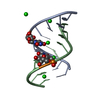
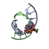
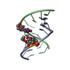
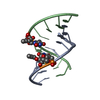
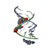
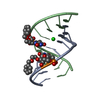
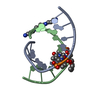
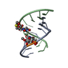
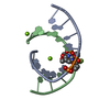
| タイトル | Crystal structure of the A-DNA GCGTAT*CGC with a 2'-O-[2-(trifluoro)ethyl] Thymidine (T*) |
|---|
 要素 要素 | 5'-D(*GP*CP*GP*TP*AP*(TFE)P*AP*CP*GP*C)-3')  キーワード キーワード | DNA / A-DNA / O2'-modification / decamer |
|---|
| 機能・相同性 | SPERMINE / DNA 機能・相同性情報 機能・相同性情報 |
|---|
| 手法 |  X線回折 / 分子置換 / 解像度: 1.5 Å X線回折 / 分子置換 / 解像度: 1.5 Å |
|---|
 データ登録者 データ登録者 | Egli, M. / Minasov, G. / Tereshko, V. / Pallan, P.S. / Teplova, M. / Inamati, G.B. / Lesnik, E.A. / Owens, S.R. / Ross, B.S. / Prakash, T.P. / Manoharan, M. |
|---|
 引用 引用 | ジャーナル: Biochemistry / 年: 2005
タイトル: Probing the Influence of Stereoelectronic Effects on the Biophysical Properties of Oligonucleotides: Comprehensive Analysis of the RNA Affinity, Nuclease Resistance, and Crystal ...タイトル: Probing the Influence of Stereoelectronic Effects on the Biophysical Properties of Oligonucleotides: Comprehensive Analysis of the RNA Affinity, Nuclease Resistance, and Crystal Structure of Ten 2'-O-Ribonucleic Acid Modifications.
著者: Egli, M. / Minasov, G. / Tereshko, V. / Pallan, P.S. / Teplova, M. / Inamati, G.B. / Lesnik, E.A. / Owens, S.R. / Ross, B.S. / Prakash, T.P. / Manoharan, M. |
|---|
| 履歴 | | 登録 | 2004年12月13日 | 登録サイト: RCSB / 処理サイト: RCSB |
|---|
| 改定 1.0 | 2005年6月28日 | Provider: repository / タイプ: Initial release |
|---|
| 改定 1.1 | 2008年4月30日 | Group: Version format compliance |
|---|
| 改定 1.2 | 2011年7月13日 | Group: Version format compliance |
|---|
| 改定 1.3 | 2023年8月23日 | Group: Data collection / Database references ...Data collection / Database references / Derived calculations / Refinement description
カテゴリ: chem_comp_atom / chem_comp_bond ...chem_comp_atom / chem_comp_bond / database_2 / pdbx_initial_refinement_model / pdbx_struct_conn_angle / struct_conn / struct_site
Item: _database_2.pdbx_DOI / _database_2.pdbx_database_accession ..._database_2.pdbx_DOI / _database_2.pdbx_database_accession / _pdbx_struct_conn_angle.ptnr1_auth_asym_id / _pdbx_struct_conn_angle.ptnr1_auth_seq_id / _pdbx_struct_conn_angle.ptnr1_label_alt_id / _pdbx_struct_conn_angle.ptnr1_label_asym_id / _pdbx_struct_conn_angle.ptnr3_auth_asym_id / _pdbx_struct_conn_angle.ptnr3_auth_seq_id / _pdbx_struct_conn_angle.ptnr3_label_alt_id / _pdbx_struct_conn_angle.ptnr3_label_asym_id / _pdbx_struct_conn_angle.value / _struct_conn.conn_type_id / _struct_conn.id / _struct_conn.pdbx_dist_value / _struct_conn.pdbx_leaving_atom_flag / _struct_conn.pdbx_ptnr2_label_alt_id / _struct_conn.ptnr1_auth_asym_id / _struct_conn.ptnr1_auth_comp_id / _struct_conn.ptnr1_auth_seq_id / _struct_conn.ptnr1_label_asym_id / _struct_conn.ptnr1_label_atom_id / _struct_conn.ptnr1_label_comp_id / _struct_conn.ptnr1_label_seq_id / _struct_conn.ptnr2_auth_asym_id / _struct_conn.ptnr2_auth_comp_id / _struct_conn.ptnr2_auth_seq_id / _struct_conn.ptnr2_label_asym_id / _struct_conn.ptnr2_label_atom_id / _struct_conn.ptnr2_label_comp_id / _struct_conn.ptnr2_label_seq_id / _struct_site.pdbx_auth_asym_id / _struct_site.pdbx_auth_comp_id / _struct_site.pdbx_auth_seq_id |
|---|
|
|---|
|
|---|
 ムービー
ムービー コントローラー


 データを開く
 基本情報
基本情報 構造の表示
構造の表示 ダウンロードとリンク
 その他のダウンロード
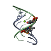


 PDBj
PDBj


 集合体
集合体

 分子量: 24.305 Da / 分子数: 1 / 由来タイプ: 合成 / 式: Mg
 分子量: 202.340 Da / 分子数: 1 / 由来タイプ: 合成 / 式: C10H26N4
 分子量: 18.015 Da / 分子数: 111 / 由来タイプ: 天然 / 式: H2O
 試料調製
試料調製 解析
解析