 Movie
Movie Controller

[English] 日本語
 Yorodumi
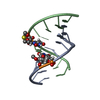
- PDB-1ma8: A-DNA decamer GCGTA(UMS)ACGC with incorporated 2'-methylseleno-uridine -
+ Open data

- Basic information
| Entry | Database: PDB / ID: 1ma8 | ||||||||||||||||||
|---|---|---|---|---|---|---|---|---|---|---|---|---|---|---|---|---|---|---|---|
| Title | A-DNA decamer GCGTA(UMS)ACGC with incorporated 2'-methylseleno-uridine | ||||||||||||||||||
 Components Components | 5'-D(* Keywords KeywordsDNA / 2'-methylseleno-uridine | Function / homology | DNA |  Function and homology information Function and homology informationMethod |  X-RAY DIFFRACTION / SYNCHROTRON / MAD / Resolution: 1.3 Å X-RAY DIFFRACTION / SYNCHROTRON / MAD / Resolution: 1.3 Å  Authors AuthorsTeplova, M. / Wilds, C.J. / Wawrzak, Z. / Tereshko, V. / Du, Q. / Carrasco, N. / Huang, Z. / Egli, M. |  CitationJournal: BIOCHIMIE / Year: 2002 CitationJournal: BIOCHIMIE / Year: 2002Title: Covalent incorporation of selenium into oligonucleotides for X-ray crystal structure determination via MAD: proof of principle Authors: Teplova, M. / Wilds, C.J. / Wawrzak, Z. / Tereshko, V. / Du, Q. / Carrasco, N. / Huang, Z. / Egli, M. History |
|
- Structure visualization
| Structure viewer | Molecule: MolmilJmol/JSmol |
|---|
- Downloads & links
-Download
| PDBx/mmCIF format | 1ma8.cif.gz | 39.6 KB | Display | PDBx/mmCIF format |
|---|---|---|---|---|
| PDB format | pdb1ma8.ent.gz | 28.6 KB | Display | PDB format |
| PDBx/mmJSON format | 1ma8.json.gz | Tree view | PDBx/mmJSON format | |
| Others |  Other downloads Other downloads |
-Validation report
| Arichive directory | https://data.pdbj.org/pub/pdb/validation_reports/ma/1ma8ftp://data.pdbj.org/pub/pdb/validation_reports/ma/1ma8 | HTTPS FTP |
|---|
-Related structure data
| Similar structure data |
|---|


-Links
 PDBj
PDBj

- Assembly
| Deposited unit | 
| ||||||||
|---|---|---|---|---|---|---|---|---|---|
| 1 |
| ||||||||
| Unit cell |
|
-Components
| #1: DNA chain | Mass: 3123.965 Da / Num. of mol.: 2 / Source method: obtained synthetically / Details: U-Se-modified DNA duplex #2: Water | ChemComp-HOH / |  Mass: 18.015 Da / Num. of mol.: 141 / Source method: isolated from a natural source / Formula: H2O Mass: 18.015 Da / Num. of mol.: 141 / Source method: isolated from a natural source / Formula: H2O |
|---|
-Experimental details
-Experiment
| Experiment | Method: X-RAY DIFFRACTION / Number of used crystals: 1 |
|---|
- Sample preparation
| Crystal | Density Matthews: 1.88 Å3/Da / Density % sol: 34.63 % | ||||||||||||||||||||||||||||||||||||||||||||||||||||||||
|---|---|---|---|---|---|---|---|---|---|---|---|---|---|---|---|---|---|---|---|---|---|---|---|---|---|---|---|---|---|---|---|---|---|---|---|---|---|---|---|---|---|---|---|---|---|---|---|---|---|---|---|---|---|---|---|---|---|
| Crystal grow | Temperature: 298 K / Method: vapor diffusion, hanging drop / pH: 7 Details: MPD, spermine tetrahydrochloride, potassium chloride, sodium chloride, sodium cacodylate, pH 7.0, VAPOR DIFFUSION, HANGING DROP, temperature 298K | ||||||||||||||||||||||||||||||||||||||||||||||||||||||||
| Components of the solutions |
| ||||||||||||||||||||||||||||||||||||||||||||||||||||||||
| Crystal grow | *PLUS | ||||||||||||||||||||||||||||||||||||||||||||||||||||||||
| Components of the solutions | *PLUS
|
-Data collection
| Diffraction | Mean temperature: 100 K |
|---|---|
| Diffraction source | Source: SYNCHROTRON / Site: APS  / Beamline: 5ID-B / Beamline: 5ID-B |
| Detector | Type: MARRESEARCH / Detector: CCD / Date: May 30, 2001 |
| Radiation | Protocol: MAD / Monochromatic (M) / Laue (L): M / Scattering type: x-ray |
| Radiation wavelength | Relative weight: 1 |
| Reflection | Resolution: 1.3→20 Å / Num. obs: 23332 / % possible obs: 99.8 % / Redundancy: 6.9 % / Rsym value: 0.049 |
| Reflection shell | Resolution: 1.3→1.35 Å / Rsym value: 0.263 / % possible all: 100 |
| Reflection | *PLUS Lowest resolution: 20 Å / Rmerge(I) obs: 0.049 |
| Reflection shell | *PLUS % possible obs: 100 % / Rmerge(I) obs: 0.263 |
- Processing
| Software |
| ||||||||||||||||
|---|---|---|---|---|---|---|---|---|---|---|---|---|---|---|---|---|---|
| Refinement | Method to determine structure: MAD / Resolution: 1.3→10 Å / σ(I): 4
| ||||||||||||||||
| Refinement step | Cycle: LAST / Resolution: 1.3→10 Å
| ||||||||||||||||
| Refine LS restraints |
| ||||||||||||||||
| Software | *PLUS Name: SHELXL / Version: 97 / Classification: refinement | ||||||||||||||||
| Refinement | *PLUS Lowest resolution: 10 Å / Num. reflection all: 23279 / % reflection Rfree: 5 % / Rfactor obs: 0.154 | ||||||||||||||||
| Solvent computation | *PLUS | ||||||||||||||||
| Displacement parameters | *PLUS |
