 Movie
Movie Controller

[English] 日本語
 Yorodumi
- PDB-6s7d: Self-complementary duplex DNA containing an internucleoside phosp... -
+ Open data

- Basic information
| Entry | Database: PDB / ID: 6s7d | ||||||||||||||||||||||||||||
|---|---|---|---|---|---|---|---|---|---|---|---|---|---|---|---|---|---|---|---|---|---|---|---|---|---|---|---|---|---|
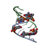
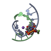
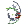
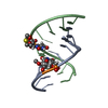
| Title | Self-complementary duplex DNA containing an internucleoside phosphoroselenolate | ||||||||||||||||||||||||||||
 Components Components | DNA (5'-D(* Keywords KeywordsDNA / Modified Phasing Duplex Phosphoroselenolate | Function / homology | : / SPERMINE / DNA |  Function and homology information Function and homology informationBiological species | synthetic construct (others) | Method |  X-RAY DIFFRACTION / SYNCHROTRON / SAD / Resolution: 1.45 Å X-RAY DIFFRACTION / SYNCHROTRON / SAD / Resolution: 1.45 Å  Authors AuthorsConlon, P.F. / Steinhogl, J. / Vyle, J.S. / Hall, J.P. | Funding support | |  United Kingdom, 1items United Kingdom, 1items
 CitationJournal: Chem Sci / Year: 2019 CitationJournal: Chem Sci / Year: 2019Title: Solid-phase synthesis and structural characterisation of phosphoroselenolate-modified DNA: a backbone analogue which does not impose conformational bias and facilitates SAD X-ray crystallography. Authors: Conlon, P.F. / Eguaogie, O. / Wilson, J.J. / Sweet, J.S.T. / Steinhoegl, J. / Englert, K. / Hancox, O.G.A. / Law, C.J. / Allman, S.A. / Tucker, J.H.R. / Hall, J.P. / Vyle, J.S. History |
|
- Structure visualization
| Structure viewer | Molecule: MolmilJmol/JSmol |
|---|
- Downloads & links
-Download
| PDBx/mmCIF format | 6s7d.cif.gz | 45.6 KB | Display | PDBx/mmCIF format |
|---|---|---|---|---|
| PDB format | pdb6s7d.ent.gz | 30.8 KB | Display | PDB format |
| PDBx/mmJSON format | 6s7d.json.gz | Tree view | PDBx/mmJSON format | |
| Others |  Other downloads Other downloads |
-Validation report
| Arichive directory | https://data.pdbj.org/pub/pdb/validation_reports/s7/6s7dftp://data.pdbj.org/pub/pdb/validation_reports/s7/6s7d | HTTPS FTP |
|---|
-Related structure data
| Similar structure data |
|---|


-Links
 PDBj
PDBj


- Assembly
| Deposited unit | 
| ||||||||||||
|---|---|---|---|---|---|---|---|---|---|---|---|---|---|
| 1 |
| ||||||||||||
| Unit cell |
|
-Components
-DNA chain , 1 types, 2 molecules AB
| #1: DNA chain | Mass: 3092.955 Da / Num. of mol.: 2 / Source method: obtained synthetically / Source: (synth.) synthetic construct (others) |
|---|
-Non-polymers , 5 types, 66 molecules 


| #2: Chemical |  Mass: 137.327 Da / Num. of mol.: 2 / Source method: obtained synthetically / Formula: Ba Mass: 137.327 Da / Num. of mol.: 2 / Source method: obtained synthetically / Formula: Ba#3: Chemical |  Mass: 22.990 Da / Num. of mol.: 2 / Source method: obtained synthetically / Formula: Na Mass: 22.990 Da / Num. of mol.: 2 / Source method: obtained synthetically / Formula: Na#4: Chemical | ChemComp-CL / |  Mass: 35.453 Da / Num. of mol.: 1 / Source method: obtained synthetically / Formula: Cl Mass: 35.453 Da / Num. of mol.: 1 / Source method: obtained synthetically / Formula: Cl#5: Chemical | ChemComp-SPM / |  Mass: 202.340 Da / Num. of mol.: 1 / Source method: obtained synthetically / Formula: C10H26N4 Mass: 202.340 Da / Num. of mol.: 1 / Source method: obtained synthetically / Formula: C10H26N4#6: Water | ChemComp-HOH / | Mass: 18.015 Da / Num. of mol.: 60 / Source method: isolated from a natural source / Formula: H2O |
|---|
-Details
| Has ligand of interest | Y |
|---|
-Experimental details
-Experiment
| Experiment | Method: X-RAY DIFFRACTION / Number of used crystals: 1 |
|---|
- Sample preparation
| Crystal | Density Matthews: 3.14 Å3/Da / Density % sol: 60.8 % |
|---|---|
| Crystal grow | Temperature: 291 K / Method: vapor diffusion, sitting drop / pH: 6 Details: The crystallization solution contained 1 uL of 2 mM oligonucleotide and 6 uL of a solution containing 10% (v/v) 2-methyl-2,4-pentanediol, 40 mM Na-cacodylate pH 6, 12 mM Spermine tetra-HCl, ...Details: The crystallization solution contained 1 uL of 2 mM oligonucleotide and 6 uL of a solution containing 10% (v/v) 2-methyl-2,4-pentanediol, 40 mM Na-cacodylate pH 6, 12 mM Spermine tetra-HCl, 80 mM NaCl and 20 mM BaCl2. This was equilibrated against 100 uL of 35% (v/v) 2-methyl-2,4-pentanediol. Crystals grew in approximately 1-2 weeks and were grown using the sitting-drop method at 291 K. |
-Data collection
| Diffraction | Mean temperature: 100 K / Serial crystal experiment: N |
|---|---|
| Diffraction source | Source: SYNCHROTRON / Site: Diamond / Beamline: I03 / Wavelength: 0.9596 Å |
| Detector | Type: DECTRIS PILATUS3 6M / Detector: PIXEL / Date: Feb 18, 2019 |
| Radiation | Monochromator: Dual Si(111) / Protocol: SINGLE WAVELENGTH / Monochromatic (M) / Laue (L): M / Scattering type: x-ray |
| Radiation wavelength | Wavelength: 0.9596 Å / Relative weight: 1 |
| Reflection | Resolution: 1.45→25.57 Å / Num. obs: 12548 / % possible obs: 98.4 % / Observed criterion σ(F): -3 / Redundancy: 3 % / CC1/2: 0.998 / Rmerge(I) obs: 0.05 / Rpim(I) all: 0.032 / Rrim(I) all: 0.06 / Net I/σ(I): 12.5 |
| Reflection shell | Resolution: 1.45→1.47 Å / Redundancy: 2.9 % / Rmerge(I) obs: 0.556 / Num. unique obs: 625 / CC1/2: 0.741 / Rpim(I) all: 0.375 / Rrim(I) all: 0.674 / % possible all: 98.7 |
- Processing
| Software |
| |||||||||||||||||||||||||||||||||||
|---|---|---|---|---|---|---|---|---|---|---|---|---|---|---|---|---|---|---|---|---|---|---|---|---|---|---|---|---|---|---|---|---|---|---|---|---|
| Refinement | Method to determine structure: SAD / Resolution: 1.45→25.57 Å / Cross valid method: FREE R-VALUE / σ(F): 3.79 / Phase error: 25.2935
| |||||||||||||||||||||||||||||||||||
| Solvent computation | Shrinkage radii: 0.9 Å / VDW probe radii: 1.11 Å | |||||||||||||||||||||||||||||||||||
| Displacement parameters | Biso mean: 25.54 Å2 | |||||||||||||||||||||||||||||||||||
| Refinement step | Cycle: LAST / Resolution: 1.45→25.57 Å
| |||||||||||||||||||||||||||||||||||
| Refine LS restraints |
| |||||||||||||||||||||||||||||||||||
| LS refinement shell |
|
