 Movie
Movie Controller

[English] 日本語
 Yorodumi
- PDB-1xye: T-to-THigh Transitions in Human Hemoglobin: alpha Y42A deoxy low salt -
+ Open data

- Basic information
| Entry | Database: PDB / ID: 1xye | ||||||
|---|---|---|---|---|---|---|---|
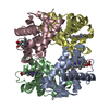
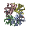
| Title | T-to-THigh Transitions in Human Hemoglobin: alpha Y42A deoxy low salt | ||||||
 Components Components |
| ||||||
 Keywords Keywords | TRANSPORT PROTEIN / hemoglobin mutant / globin | ||||||
| Function / homology |  Function and homology information Function and homology informationnitric oxide transport / cellular oxidant detoxification / hemoglobin binding / hemoglobin alpha binding / haptoglobin-hemoglobin complex / organic acid binding / renal absorption / hemoglobin complex / oxygen transport / Scavenging of heme from plasma ...nitric oxide transport / cellular oxidant detoxification / hemoglobin binding / hemoglobin alpha binding / haptoglobin-hemoglobin complex / organic acid binding / renal absorption / hemoglobin complex / oxygen transport / Scavenging of heme from plasma / endocytic vesicle lumen / blood vessel diameter maintenance / hydrogen peroxide catabolic process / oxygen carrier activity / regulation of blood pressure / Late endosomal microautophagy / Heme signaling / carbon dioxide transport / Erythrocytes take up oxygen and release carbon dioxide / response to hydrogen peroxide / Erythrocytes take up carbon dioxide and release oxygen / Cytoprotection by HMOX1 / platelet aggregation / oxygen binding / Chaperone Mediated Autophagy / positive regulation of nitric oxide biosynthetic process / Factors involved in megakaryocyte development and platelet production / tertiary granule lumen / ficolin-1-rich granule lumen / blood microparticle / iron ion binding / heme binding / Neutrophil degranulation / extracellular space / extracellular exosome / extracellular region / membrane / metal ion binding / cytosol Similarity search - Function | ||||||
| Biological species |  Homo sapiens (human) Homo sapiens (human) | ||||||
| Method |  X-RAY DIFFRACTION / MOLECULAR REPLACEMENT / Resolution: 2.13 Å X-RAY DIFFRACTION / MOLECULAR REPLACEMENT / Resolution: 2.13 Å | ||||||
 Authors Authors | Kavanaugh, J.S. / Rogers, P.H. / Arnone, A. / Hui, H.L. / Wierzba, A. / DeYoung, A. / Kwiatkowski, L.D. / Noble, R.W. / Juszczak, L.J. / Peterson, E.S. / Friedman, J.M. | ||||||
 Citation Citation | Journal: Biochemistry / Year: 2005 Title: Intersubunit interactions associated with tyr42alpha stabilize the quaternary-T tetramer but are not major quaternary constraints in deoxyhemoglobin Authors: Kavanaugh, J.S. / Rogers, P.H. / Arnone, A. / Hui, H.L. / Wierzba, A. / Deyoung, A. / Kwiatkowski, L.D. / Noble, R.W. / Juszczak, L.J. / Peterson, E.S. / Friedman, J.M. #1: Journal: To be PublishedTitle: Crystallographic Evidence for a New Ensemble of Ligand-Induced Allosteric Transitions in Hemoglobin: The T-to-THigh Quaternary Transitions Authors: Kavanaugh, J.S. / Rogers, P.H. / Arnone, A. | ||||||
| History |
|
- Structure visualization
| Structure viewer | Molecule: MolmilJmol/JSmol |
|---|
- Downloads & links
-Download
| PDBx/mmCIF format | 1xye.cif.gz | 126.5 KB | Display | PDBx/mmCIF format |
|---|---|---|---|---|
| PDB format | pdb1xye.ent.gz | 100 KB | Display | PDB format |
| PDBx/mmJSON format | 1xye.json.gz | Tree view | PDBx/mmJSON format | |
| Others |  Other downloads Other downloads |
-Validation report
| Summary document | 1xye_validation.pdf.gz | 1.8 MB | Display | wwPDB validaton report |
|---|---|---|---|---|
| Full document | 1xye_full_validation.pdf.gz | 1.8 MB | Display | |
| Data in XML | 1xye_validation.xml.gz | 26.6 KB | Display | |
| Data in CIF | 1xye_validation.cif.gz | 35.5 KB | Display | |
| Arichive directory | https://data.pdbj.org/pub/pdb/validation_reports/xy/1xyeftp://data.pdbj.org/pub/pdb/validation_reports/xy/1xye | HTTPS FTP |
-Related structure data
| Related structure data |  1xz2C  1xz4C  1rq3S C: citing same article ( S: Starting model for refinement |
|---|---|
| Similar structure data |


-Links
 PDBj
PDBj


- Assembly
| Deposited unit | 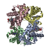
| ||||||||
|---|---|---|---|---|---|---|---|---|---|
| 1 |
| ||||||||
| Unit cell |
| ||||||||
| Details | the crystallographic asymmetric unit in this entry is an alpha2beta2 tetramer. the biological unit is an alpha2beta2 tetramer. The biological unit and crystallographic asymmetric unit are equivalent |
-Components
| #1: Protein | Mass: 15090.321 Da / Num. of mol.: 2 / Mutation: V1M, Y42A Source method: isolated from a genetically manipulated source Source: (gene. exp.) Homo sapiens (human) / Gene: HBA1 / Production host:  #2: Protein | Mass: 15890.198 Da / Num. of mol.: 2 / Source method: isolated from a natural source / Source: (natural) Homo sapiens (human) / Tissue: blood / References: UniProt: P68871#3: Chemical | ChemComp-HEM /   Mass: 616.487 Da / Num. of mol.: 4 / Source method: obtained synthetically / Formula: C34H32FeN4O4 Mass: 616.487 Da / Num. of mol.: 4 / Source method: obtained synthetically / Formula: C34H32FeN4O4#4: Water | ChemComp-HOH / |  Mass: 18.015 Da / Num. of mol.: 171 / Source method: isolated from a natural source / Formula: H2O Mass: 18.015 Da / Num. of mol.: 171 / Source method: isolated from a natural source / Formula: H2O |
|---|
-Experimental details
-Experiment
| Experiment | Method: X-RAY DIFFRACTION / Number of used crystals: 1 |
|---|
- Sample preparation
| Crystal | Density Matthews: 2.57 Å3/Da / Density % sol: 52.19 % |
|---|---|
| Crystal grow | Temperature: 298 K / Method: batch / pH: 7 Details: 10% PEG 6000, 10 mM potassium phosphate, 100 mM potassium chloride, 3 mM sodium dithionite, 10 mg/ml Hb, pH 7.0, batch, temperature 298K |
-Data collection
| Diffraction | Mean temperature: 298 K |
|---|---|
| Diffraction source | Source: ROTATING ANODE / Type: RIGAKU RU200 / Wavelength: 1.5418 Å |
| Detector | Type: SDMS / Detector: AREA DETECTOR / Date: Nov 11, 1995 / Details: graphite |
| Radiation | Monochromator: graphite / Protocol: SINGLE WAVELENGTH / Monochromatic (M) / Laue (L): M / Scattering type: x-ray |
| Radiation wavelength | Wavelength: 1.5418 Å / Relative weight: 1 |
| Reflection | Highest resolution: 2.13 Å / Num. all: 36379 / Num. obs: 36379 / % possible obs: 97.9 % / Observed criterion σ(F): 0 / Observed criterion σ(I): 0 / Redundancy: 6.7 % / Rmerge(I) obs: 0.059 / Net I/σ(I): 5.4 |
| Reflection shell | Resolution: 2.13→2.29 Å / Redundancy: 3.1 % / Rmerge(I) obs: 0.185 / Mean I/σ(I) obs: 1.3 / Num. unique all: 6440 / % possible all: 90.2 |
- Processing
| Software |
| |||||||||||||||||||||||||||||||||||||||||||||||||||||||||||||||||||||||||||||||||||||||||||||||||||||||||
|---|---|---|---|---|---|---|---|---|---|---|---|---|---|---|---|---|---|---|---|---|---|---|---|---|---|---|---|---|---|---|---|---|---|---|---|---|---|---|---|---|---|---|---|---|---|---|---|---|---|---|---|---|---|---|---|---|---|---|---|---|---|---|---|---|---|---|---|---|---|---|---|---|---|---|---|---|---|---|---|---|---|---|---|---|---|---|---|---|---|---|---|---|---|---|---|---|---|---|---|---|---|---|---|---|---|---|
| Refinement | Method to determine structure: MOLECULAR REPLACEMENT Starting model: pdb entry 1RQ3 Resolution: 2.13→10 Å / Cor.coef. Fo:Fc: 0.942 / Cor.coef. Fo:Fc free: 0.904 / SU B: 9.493 / SU ML: 0.238 / Cross valid method: THROUGHOUT and Local R-free / σ(F): 0 / ESU R: 0.259 / ESU R Free: 0.207 / Stereochemistry target values: MAXIMUM LIKELIHOOD / Details: HYDROGENS HAVE BEEN ADDED IN THE RIDING POSITIONS
| |||||||||||||||||||||||||||||||||||||||||||||||||||||||||||||||||||||||||||||||||||||||||||||||||||||||||
| Solvent computation | Ion probe radii: 0.8 Å / Shrinkage radii: 0.8 Å / VDW probe radii: 1.4 Å / Solvent model: BABINET MODEL WITH MASK | |||||||||||||||||||||||||||||||||||||||||||||||||||||||||||||||||||||||||||||||||||||||||||||||||||||||||
| Displacement parameters | Biso mean: 19.671 Å2
| |||||||||||||||||||||||||||||||||||||||||||||||||||||||||||||||||||||||||||||||||||||||||||||||||||||||||
| Refinement step | Cycle: LAST / Resolution: 2.13→10 Å
| |||||||||||||||||||||||||||||||||||||||||||||||||||||||||||||||||||||||||||||||||||||||||||||||||||||||||
| Refine LS restraints |
| |||||||||||||||||||||||||||||||||||||||||||||||||||||||||||||||||||||||||||||||||||||||||||||||||||||||||
| LS refinement shell | Resolution: 2.13→2.292 Å / Total num. of bins used: 7 /
|
