 Movie
Movie Controller

[English] 日本語
 Yorodumi
- PDB-1xqm: Variations on the GFP chromophore scaffold: A fragmented 5-member... -
+ Open data

- Basic information
| Entry | Database: PDB / ID: 1xqm | ||||||
|---|---|---|---|---|---|---|---|
| Title | Variations on the GFP chromophore scaffold: A fragmented 5-membered heterocycle revealed in the 2.1A crystal structure of a non-fluorescent chromoprotein | ||||||
 Components Components | kindling fluorescent protein | ||||||
 Keywords Keywords |  LUMINESCENT PROTEIN LUMINESCENT PROTEIN | ||||||
| Function / homology |  Function and homology information Function and homology information | ||||||
| Method | X-RAY DIFFRACTION / MOLECULAR REPLACEMENT / Resolution: 2.1 Å | ||||||
 Authors Authors | Wilmann, P.G. / Petersen, J. / Devenish, R.J. / Prescott, M. / Rossjohn, J. | ||||||
 Citation Citation | Journal: J.Biol.Chem. / Year: 2005 Title: Variations on the GFP chromophore: A polypeptide fragmentation within the chromophore revealed in the 2.1-A crystal structure of a nonfluorescent chromoprotein from Anemonia sulcata Authors: Wilmann, P.G. / Petersen, J. / Devenish, R.J. / Prescott, M. / Rossjohn, J. | ||||||
| History |
|
- Structure visualization
| Structure viewer | Molecule: MolmilJmol/JSmol |
|---|
- Downloads & links
-Download
| PDBx/mmCIF format | 1xqm.cif.gz | 55.7 KB | Display | PDBx/mmCIF format |
|---|---|---|---|---|
| PDB format | pdb1xqm.ent.gz | 44.2 KB | Display | PDB format |
| PDBx/mmJSON format | 1xqm.json.gz | Tree view | PDBx/mmJSON format | |
| Others |  Other downloads Other downloads |
-Validation report
| Arichive directory | https://data.pdbj.org/pub/pdb/validation_reports/xq/1xqmftp://data.pdbj.org/pub/pdb/validation_reports/xq/1xqm | HTTPS FTP |
|---|
-Related structure data
| Similar structure data |
|---|


-Links
 PDBj
PDBj


- Assembly
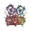
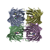
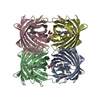
| Deposited unit | 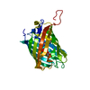
| ||||||||
|---|---|---|---|---|---|---|---|---|---|
| 1 | 
| ||||||||
| Unit cell |
| ||||||||
| Components on special symmetry positions |
|
-Components
| #1: Protein | Mass: 25838.486 Da / Num. of mol.: 1 / Source method: obtained synthetically Details: synthetic construct, It is based on the sequence from Anemonia sulcata. References: GenBank: 28629493, UniProt: Q9GZ28*PLUS |
|---|---|
| #2: Chemical | ChemComp-ACY / Acetic acid  Mass: 60.052 Da / Num. of mol.: 1 / Source method: obtained synthetically / Formula: C2H4O2 Mass: 60.052 Da / Num. of mol.: 1 / Source method: obtained synthetically / Formula: C2H4O2 |
| #3: Water | ChemComp-HOH / Water Mass: 18.015 Da / Num. of mol.: 130 / Source method: isolated from a natural source / Formula: H2O Mass: 18.015 Da / Num. of mol.: 130 / Source method: isolated from a natural source / Formula: H2O |
| Sequence details | MET A 63, TYR A 64 AND GLY A 65 ARE MODIFIED TO MAKE CHROMOPHOR |
-Experimental details
-Experiment
| Experiment | Method: X-RAY DIFFRACTION / Number of used crystals: 1 |
|---|
- Sample preparation
| Crystal | Density Matthews: 3.9 Å3/Da / Density % sol: 68.3 % |
|---|---|
| Crystal grow | Temperature: 293 K / Method: vapor diffusion, hanging drop / pH: 5.7 Details: PEG3350, Ammonium Acetate, pH 5.7, VAPOR DIFFUSION, HANGING DROP, temperature 293K |
-Data collection
| Diffraction | Mean temperature: 100 K |
|---|---|
| Diffraction source | Source: ROTATING ANODE / Type: RIGAKU / Wavelength: 1.5418 Å |
| Detector | Type: RIGAKU RAXIS IV / Detector: IMAGE PLATE / Date: Nov 20, 2003 |
| Radiation | Monochromator: osmic mirrors / Protocol: SINGLE WAVELENGTH / Monochromatic (M) / Laue (L): M / Scattering type: x-ray |
| Radiation wavelength | Wavelength: 1.5418 Å / Relative weight: 1 |
| Reflection | Resolution: 2.1→50 Å / Num. all: 21669 / Num. obs: 21515 / % possible obs: 98.6 % / Observed criterion σ(F): 1 / Observed criterion σ(I): 1 |
- Processing
| Software | Name: CNS / Classification: refinement | |||||||||||||||
|---|---|---|---|---|---|---|---|---|---|---|---|---|---|---|---|---|
| Refinement | Method to determine structure: MOLECULAR REPLACEMENT / Resolution: 2.1→50 Å / σ(F): 2
| |||||||||||||||
| Refinement step | Cycle: LAST / Resolution: 2.1→50 Å
|
