 Movie
Movie Controller


+ Open data

- Basic information
| Entry | Database: PDB / ID: 1xec | |||||||||
|---|---|---|---|---|---|---|---|---|---|---|
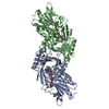
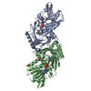
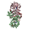
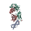
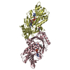
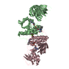
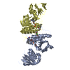
| Title | Dimeric bovine tissue-extracted decorin, crystal form 2 | |||||||||
 Components Components | Decorin | |||||||||
 Keywords Keywords | STRUCTURAL PROTEIN / PROTEOGLYCAN / LEUCINE-RICH REPEAT | |||||||||
| Function / homology |  Function and homology information Function and homology informationGlycosaminoglycan-protein linkage region biosynthesis / CS-GAG biosynthesis / DS-GAG biosynthesis / CS/DS degradation / negative regulation of collagen fibril organization / collagen fibril binding / Degradation of the extracellular matrix / ECM proteoglycans / negative regulation of vascular endothelial growth factor signaling pathway / glycosaminoglycan binding ...Glycosaminoglycan-protein linkage region biosynthesis / CS-GAG biosynthesis / DS-GAG biosynthesis / CS/DS degradation / negative regulation of collagen fibril organization / collagen fibril binding / Degradation of the extracellular matrix / ECM proteoglycans / negative regulation of vascular endothelial growth factor signaling pathway / glycosaminoglycan binding / negative regulation of endothelial cell migration / positive regulation of mitochondrial fission / positive regulation of mitochondrial depolarization / positive regulation of macroautophagy / negative regulation of angiogenesis / extracellular matrix / positive regulation of phosphatidylinositol 3-kinase/protein kinase B signal transduction / protein homodimerization activity / positive regulation of transcription by RNA polymerase II / extracellular space Similarity search - Function | |||||||||
| Biological species |  | |||||||||
| Method |  X-RAY DIFFRACTION / SYNCHROTRON / MOLECULAR REPLACEMENT / Resolution: 2.3 Å X-RAY DIFFRACTION / SYNCHROTRON / MOLECULAR REPLACEMENT / Resolution: 2.3 Å | |||||||||
 Authors Authors | Scott, P.G. / McEwan, P.A. / Dodd, C.M. / Bergmann, E.M. / Bishop, P.N. / Bella, J. | |||||||||
 Citation Citation | Journal: Proc.Natl.Acad.Sci.USA / Year: 2004 Title: Crystal structure of the dimeric protein core of decorin, the archetypal small leucine-rich repeat proteoglycan Authors: Scott, P.G. / McEwan, P.A. / Dodd, C.M. / Bergmann, E.M. / Bishop, P.N. / Bella, J. #1: Journal: J.Biol.Chem. / Year: 2003Title: Light and X-ray scattering show decorin to be a dimer in solution Authors: Scott, P.G. / Grossmann, J.G. / Dodd, C.M. / Sheehan, J.K. / Bishop, P.N. | |||||||||
| History |
| |||||||||
| Remark 290 | CRYSTALLOGRAPHIC SYMMETRY SYMMETRY OPERATORS FOR SPACE GROUP: P 21 21 21 SYMOP SYMMETRY NNNMMM ... CRYSTALLOGRAPHIC SYMMETRY SYMMETRY OPERATORS FOR SPACE GROUP: P 21 21 21 SYMOP SYMMETRY NNNMMM OPERATOR 1555 X,Y,Z 2555 1/2-X,-Y,1/2+Z 3555 -X,1/2+Y,1/2-Z 4555 1/2+X,1/2-Y,-Z WHERE NNN -> OPERATOR NUMBER MMM -> TRANSLATION VECTOR CRYSTALLOGRAPHIC SYMMETRY TRANSFORMATIONS THE FOLLOWING TRANSFORMATIONS OPERATE ON THE ATOM/HETATM RECORDS IN THIS ENTRY TO PRODUCE CRYSTALLOGRAPHICALLY RELATED MOLECULES. SMTRY1 1 1.000000 0.000000 0.000000 0.00000 SMTRY2 1 0.000000 1.000000 0.000000 0.00000 SMTRY3 1 0.000000 0.000000 1.000000 0.00000 SMTRY1 2 -1.000000 0.000000 0.000000 26.35050 SMTRY2 2 0.000000 -1.000000 0.000000 0.00000 SMTRY3 2 0.000000 0.000000 1.000000 64.85600 SMTRY1 3 -1.000000 0.000000 0.000000 0.00000 SMTRY2 3 0.000000 1.000000 0.000000 60.47600 SMTRY3 3 0.000000 0.000000 -1.000000 64.85600 SMTRY1 4 1.000000 0.000000 0.000000 26.35050 SMTRY2 4 0.000000 -1.000000 0.000000 60.47600 SMTRY3 4 0.000000 0.000000 -1.000000 0.00000 REMARK: NULL |
- Structure visualization
| Structure viewer | Molecule: MolmilJmol/JSmol |
|---|
- Downloads & links
-Download
| PDBx/mmCIF format | 1xec.cif.gz | 138.3 KB | Display | PDBx/mmCIF format |
|---|---|---|---|---|
| PDB format | pdb1xec.ent.gz | 107.8 KB | Display | PDB format |
| PDBx/mmJSON format | 1xec.json.gz | Tree view | PDBx/mmJSON format | |
| Others |  Other downloads Other downloads |
-Validation report
| Arichive directory | https://data.pdbj.org/pub/pdb/validation_reports/xe/1xecftp://data.pdbj.org/pub/pdb/validation_reports/xe/1xec | HTTPS FTP |
|---|
-Related structure data
| Related structure data |  1xcdC  1xkuSC S: Starting model for refinement C: citing same article ( |
|---|---|
| Similar structure data |
-Links
 PDBj
PDBj


- Assembly
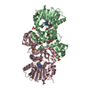
| Deposited unit | 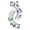
| ||||||||||
|---|---|---|---|---|---|---|---|---|---|---|---|
| 1 |
| ||||||||||
| Unit cell |
|
-Components
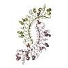
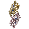
| #1: Protein | Mass: 36383.758 Da / Num. of mol.: 2 / Source method: isolated from a natural source Details: PROTEIN WAS EXTRACTED FROM CALF SKIN UNDER DENATURING CONDITIONS AND REFOLDED Source: (natural) #2: Polysaccharide | Source method: isolated from a genetically manipulated source #3: Sugar |   Type: D-saccharide, beta linking / Mass: 221.208 Da / Num. of mol.: 3 Type: D-saccharide, beta linking / Mass: 221.208 Da / Num. of mol.: 3Source method: isolated from a genetically manipulated source Formula: C8H15NO6 #4: Water | ChemComp-HOH / |  Mass: 18.015 Da / Num. of mol.: 298 / Source method: isolated from a natural source / Formula: H2O Mass: 18.015 Da / Num. of mol.: 298 / Source method: isolated from a natural source / Formula: H2OHas protein modification | Y | |
|---|
-Experimental details
-Experiment
| Experiment | Method: X-RAY DIFFRACTION / Number of used crystals: 1 |
|---|
- Sample preparation
| Crystal | Density Matthews: 2.97 Å3/Da / Density % sol: 58.2 % |
|---|---|
| Crystal grow | Temperature: 293 K / Method: vapor diffusion, hanging drop / pH: 7.75 Details: PEG 400, TRIS, OCTYL-BETA-D-GLUCOPYRANOSIDE, SODIUM AZIDE, pH 7.75, VAPOR DIFFUSION, HANGING DROP, temperature 293K |
-Data collection
| Diffraction | Mean temperature: 100 K |
|---|---|
| Diffraction source | Source: SYNCHROTRON / Site: SRS  / Beamline: PX14.2 / Wavelength: 0.978 Å / Beamline: PX14.2 / Wavelength: 0.978 Å |
| Detector | Type: ADSC QUANTUM 4 / Detector: CCD / Date: May 5, 2001 / Details: mirrors |
| Radiation | Monochromator: Si 111 CHANNEL / Protocol: SINGLE WAVELENGTH / Monochromatic (M) / Laue (L): M / Scattering type: x-ray |
| Radiation wavelength | Wavelength: 0.978 Å / Relative weight: 1 |
| Reflection | Resolution: 2.3→19.73 Å / Num. all: 37599 / Num. obs: 37599 / % possible obs: 96.7 % / Observed criterion σ(F): 0 / Observed criterion σ(I): 0 / Redundancy: 5.2 % / Biso Wilson estimate: 31.6 Å2 / Rsym value: 0.09 / Net I/σ(I): 6.3 |
| Reflection shell | Resolution: 2.3→2.44 Å / Redundancy: 4.7 % / Mean I/σ(I) obs: 2.9 / Num. unique all: 3024 / Rsym value: 0.199 / % possible all: 83.5 |
- Processing
| Software |
| ||||||||||||||||||||||||||||||||||||
|---|---|---|---|---|---|---|---|---|---|---|---|---|---|---|---|---|---|---|---|---|---|---|---|---|---|---|---|---|---|---|---|---|---|---|---|---|---|
| Refinement | Method to determine structure: MOLECULAR REPLACEMENT Starting model: PDB ENTRY 1XKU Resolution: 2.3→19.73 Å / Rfactor Rfree error: 0.004 / Data cutoff high absF: 1680211.91 / Data cutoff low absF: 0 / Isotropic thermal model: RESTRAINED / Cross valid method: THROUGHOUT / σ(F): 0 / Stereochemistry target values: Engh & Huber
| ||||||||||||||||||||||||||||||||||||
| Solvent computation | Solvent model: FLAT MODEL / Bsol: 29.5121 Å2 / ksol: 0.2985 e/Å3 | ||||||||||||||||||||||||||||||||||||
| Displacement parameters | Biso mean: 48.4 Å2
| ||||||||||||||||||||||||||||||||||||
| Refine analyze |
| ||||||||||||||||||||||||||||||||||||
| Refinement step | Cycle: LAST / Resolution: 2.3→19.73 Å
| ||||||||||||||||||||||||||||||||||||
| Refine LS restraints |
| ||||||||||||||||||||||||||||||||||||
| LS refinement shell | Resolution: 2.3→2.44 Å / Rfactor Rfree error: 0.016 / Total num. of bins used: 6
| ||||||||||||||||||||||||||||||||||||
| Xplor file |
|
