 Movie
Movie Controller


+ Open data

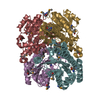
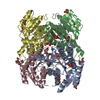
- Basic information
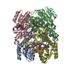
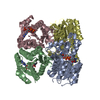
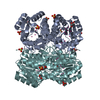
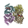
| Entry | Database: PDB / ID: 1wnt | ||||||
|---|---|---|---|---|---|---|---|
| Title | Structure of the tetrameric form of Human L-Xylulose Reductase | ||||||
 Components Components | L-xylulose reductase | ||||||
 Keywords Keywords | OXIDOREDUCTASE / 7 stranded parallel beta sheets / 6 alpha helices / Rossmann fold / dinucleotide co-enzyme binding motif | ||||||
| Function / homology |  Function and homology informationEssential pentosuria / L-xylulose reductase / L-xylulose reductase (NADPH) activity / xylulose metabolic process / glucuronate catabolic process to xylulose 5-phosphate / Formation of xylulose-5-phosphate / carbonyl reductase (NADPH) activity / NADP metabolic process / D-xylose metabolic process / oxidoreductase activity, acting on NAD(P)H, quinone or similar compound as acceptor ...Essential pentosuria / L-xylulose reductase / L-xylulose reductase (NADPH) activity / xylulose metabolic process / glucuronate catabolic process to xylulose 5-phosphate / Formation of xylulose-5-phosphate / carbonyl reductase (NADPH) activity / NADP metabolic process / D-xylose metabolic process / oxidoreductase activity, acting on NAD(P)H, quinone or similar compound as acceptor / microvillus / brush border / cytoplasmic microtubule / glucose metabolic process / positive regulation of reactive oxygen species metabolic process / extracellular exosome / identical protein binding / nucleus / plasma membrane / cytosol Function and homology informationEssential pentosuria / L-xylulose reductase / L-xylulose reductase (NADPH) activity / xylulose metabolic process / glucuronate catabolic process to xylulose 5-phosphate / Formation of xylulose-5-phosphate / carbonyl reductase (NADPH) activity / NADP metabolic process / D-xylose metabolic process / oxidoreductase activity, acting on NAD(P)H, quinone or similar compound as acceptor ...Essential pentosuria / L-xylulose reductase / L-xylulose reductase (NADPH) activity / xylulose metabolic process / glucuronate catabolic process to xylulose 5-phosphate / Formation of xylulose-5-phosphate / carbonyl reductase (NADPH) activity / NADP metabolic process / D-xylose metabolic process / oxidoreductase activity, acting on NAD(P)H, quinone or similar compound as acceptor / microvillus / brush border / cytoplasmic microtubule / glucose metabolic process / positive regulation of reactive oxygen species metabolic process / extracellular exosome / identical protein binding / nucleus / plasma membrane / cytosolSimilarity search - Function | ||||||
| Biological species |  Homo sapiens (human) Homo sapiens (human) | ||||||
| Method | X-RAY DIFFRACTION / MOLECULAR REPLACEMENT / Resolution: 2.3 Å | ||||||
 Authors Authors | El-Kabbani, O. / Carbone, V. / Darmanin, C. / Ishikura, S. / Hara, A. | ||||||
 Citation Citation | Journal: Proteins / Year: 2005 Title: Structure of the tetrameric form of human L-Xylulose reductase: Probing the inhibitor-binding site with molecular modeling and site-directed mutagenesis Authors: El-Kabbani, O. / Carbone, V. / Darmanin, C. / Ishikura, S. / Hara, A. | ||||||
| History |
|
- Structure visualization
| Structure viewer | Molecule: MolmilJmol/JSmol |
|---|
- Downloads & links
-Download
| PDBx/mmCIF format | 1wnt.cif.gz | 195.4 KB | Display | PDBx/mmCIF format |
|---|---|---|---|---|
| PDB format | pdb1wnt.ent.gz | 157.3 KB | Display | PDB format |
| PDBx/mmJSON format | 1wnt.json.gz | Tree view | PDBx/mmJSON format | |
| Others |  Other downloads Other downloads |
-Validation report
| Arichive directory | https://data.pdbj.org/pub/pdb/validation_reports/wn/1wntftp://data.pdbj.org/pub/pdb/validation_reports/wn/1wnt | HTTPS FTP |
|---|
-Related structure data
| Related structure data |  1pr9S S: Starting model for refinement |
|---|---|
| Similar structure data |


-Links
 PDBj
PDBj


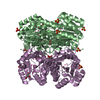
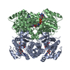
- Assembly
| Deposited unit | 
| ||||||||
|---|---|---|---|---|---|---|---|---|---|
| 1 |
| ||||||||
| Unit cell |
|
-Components
| #1: Protein | / XR / Dicarbonyl/L-xylulose reductase / Kidney dicarbonyl reductase / kiDCR / Carbonyl reductase II ...XR / Dicarbonyl/L-xylulose reductase / Kidney dicarbonyl reductase / kiDCR / Carbonyl reductase II / Sperm surface protein P34H Mass: 25941.033 Da / Num. of mol.: 4 Source method: isolated from a genetically manipulated source Source: (gene. exp.) Homo sapiens (human) / Tissue: kidney / Plasmid: pRset / Species (production host): Escherichia coli / Production host:  Escherichia coli BL21 (bacteria) / Strain (production host): BL21 / References: UniProt: Q7Z4W1, L-xylulose reductase Escherichia coli BL21 (bacteria) / Strain (production host): BL21 / References: UniProt: Q7Z4W1, L-xylulose reductase#2: Chemical | ChemComp-NAP / Nicotinamide adenine dinucleotide phosphate  Mass: 743.405 Da / Num. of mol.: 4 / Source method: obtained synthetically / Formula: C21H28N7O17P3 Mass: 743.405 Da / Num. of mol.: 4 / Source method: obtained synthetically / Formula: C21H28N7O17P3#3: Water | ChemComp-HOH / | Water Mass: 18.015 Da / Num. of mol.: 146 / Source method: isolated from a natural source / Formula: H2O Mass: 18.015 Da / Num. of mol.: 146 / Source method: isolated from a natural source / Formula: H2O |
|---|
-Experimental details
-Experiment
| Experiment | Method: X-RAY DIFFRACTION / Number of used crystals: 1 |
|---|
- Sample preparation
| Crystal | Density Matthews: 2.1 Å3/Da / Density % sol: 41 % |
|---|---|
| Crystal grow | Temperature: 295 K / Method: vapor diffusion, hanging drop / pH: 7.5 Details: PEG 4000, Sodium Citrate, Isopropanol, Urea, pH 7.5, VAPOR DIFFUSION, HANGING DROP, temperature 295.0K |
-Data collection
| Diffraction | Mean temperature: 100 K |
|---|---|
| Diffraction source | Source: ROTATING ANODE / Type: RIGAKU RU300 / Wavelength: 1.5418 Å |
| Detector | Type: RIGAKU RAXIS IV / Detector: IMAGE PLATE / Date: Aug 10, 2003 / Details: mirrors |
| Radiation | Monochromator: Capillary optics / Protocol: SINGLE WAVELENGTH / Monochromatic (M) / Laue (L): M / Scattering type: x-ray |
| Radiation wavelength | Wavelength: 1.5418 Å / Relative weight: 1 |
| Reflection | Resolution: 2.3→20 Å / Num. all: 64193 / Num. obs: 32262 / % possible obs: 92 % / Observed criterion σ(F): 4 / Observed criterion σ(I): 2 / Redundancy: 1.99 % / Rsym value: 0.09 |
| Reflection shell | Resolution: 2.3→2.38 Å / Mean I/σ(I) obs: 1.98 / Rsym value: 0.346 / % possible all: 90.9 |
- Processing
| Software |
| ||||||||||||||||||||
|---|---|---|---|---|---|---|---|---|---|---|---|---|---|---|---|---|---|---|---|---|---|
| Refinement | Method to determine structure: MOLECULAR REPLACEMENT Starting model: XR dimer (1PR9) Resolution: 2.3→12 Å / Isotropic thermal model: Isotropic / σ(F): 4 / Stereochemistry target values: Engh & Huber
| ||||||||||||||||||||
| Refine analyze | Luzzati coordinate error obs: 0.25 Å | ||||||||||||||||||||
| Refinement step | Cycle: LAST / Resolution: 2.3→12 Å
| ||||||||||||||||||||
| Refine LS restraints |
|
