 Movie
Movie Controller

[English] 日本語
 Yorodumi
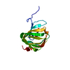
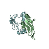
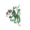
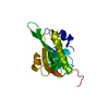
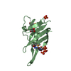
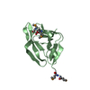
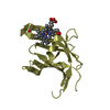
- PDB-1vfq: The Crystal Structure of Human Coactosin-like Protein at 1.9 A Re... -
+ Open data

- Basic information
| Entry | Database: PDB / ID: 1vfq | ||||||
|---|---|---|---|---|---|---|---|
| Title | The Crystal Structure of Human Coactosin-like Protein at 1.9 A Resolution | ||||||
 Components Components | Coactosin-like protein | ||||||
 Keywords Keywords |  PROTEIN BINDING / Cytoskeleton / Actin-binding protein PROTEIN BINDING / Cytoskeleton / Actin-binding protein | ||||||
| Function / homology |  Function and homology information Function and homology informationsite of polarized growth / regulation of actin filament polymerization / cortical actin cytoskeleton / defense response to fungus / actin filament binding / actin binding / secretory granule lumen / ficolin-1-rich granule lumen / Neutrophil degranulation / enzyme binding ...site of polarized growth / regulation of actin filament polymerization / cortical actin cytoskeleton / defense response to fungus / actin filament binding / actin binding / secretory granule lumen / ficolin-1-rich granule lumen / Neutrophil degranulation / enzyme binding / extracellular exosome / extracellular region / nucleus / cytosolSimilarity search - Function | ||||||
| Biological species |  Homo sapiens (human) Homo sapiens (human) | ||||||
| Method | X-RAY DIFFRACTION / MOLECULAR REPLACEMENT / Resolution: 1.9 Å | ||||||
 Authors Authors | Li, X. / Liu, Y. / Liu, X. / Lou, Z. | ||||||
 Citation Citation | Journal: PROTEIN SCI. / Year: 2004 Title: Crystal structure of human coactosin-like protein at 1.9 A resolution Authors: Li, X. / Liu, X. / Lou, Z. / Duan, X. / Wu, H. / Liu, Y. / Rao, Z. | ||||||
| History |
|
- Structure visualization
| Structure viewer | Molecule: MolmilJmol/JSmol |
|---|
- Downloads & links
-Download
| PDBx/mmCIF format | 1vfq.cif.gz | 40 KB | Display | PDBx/mmCIF format |
|---|---|---|---|---|
| PDB format | pdb1vfq.ent.gz | 27.1 KB | Display | PDB format |
| PDBx/mmJSON format | 1vfq.json.gz | Tree view | PDBx/mmJSON format | |
| Others |  Other downloads Other downloads |
-Validation report
| Arichive directory | https://data.pdbj.org/pub/pdb/validation_reports/vf/1vfqftp://data.pdbj.org/pub/pdb/validation_reports/vf/1vfq | HTTPS FTP |
|---|
-Related structure data
| Similar structure data |
|---|


-Links
 PDBj
PDBj
- Assembly
| Deposited unit | 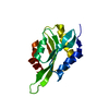
| ||||||||
|---|---|---|---|---|---|---|---|---|---|
| 1 |
| ||||||||
| Unit cell |
|
-Components
| #1: Protein | Mass: 16053.041 Da / Num. of mol.: 1 Source method: isolated from a genetically manipulated source Source: (gene. exp.) Homo sapiens (human) / Plasmid: pGEX-6p-1 / Production host:  Escherichia coli (E. coli) / References: UniProt: Q14019 Escherichia coli (E. coli) / References: UniProt: Q14019 |
|---|---|
| #2: Water | ChemComp-HOH / Water Mass: 18.015 Da / Num. of mol.: 110 / Source method: isolated from a natural source / Formula: H2O Mass: 18.015 Da / Num. of mol.: 110 / Source method: isolated from a natural source / Formula: H2O |
-Experimental details
-Experiment
| Experiment | Method: X-RAY DIFFRACTION / Number of used crystals: 1 |
|---|
- Sample preparation
| Crystal | Density Matthews: 1.6 Å3/Da / Density % sol: 24 % |
|---|---|
| Crystal grow | Temperature: 291 K / Method: vapor diffusion, hanging drop / pH: 8.5 Details: 30% PEG4000, 0.1M Tris-HCL, pH 8.5, VAPOR DIFFUSION, HANGING DROP, temperature 291K |
-Data collection
| Diffraction | Mean temperature: 100 K |
|---|---|
| Diffraction source | Source: ROTATING ANODE / Type: RIGAKU / Wavelength: 1.5418 Å |
| Detector | Type: MARRESEARCH / Detector: AREA DETECTOR / Date: Jun 17, 2003 |
| Radiation | Monochromator: osmic mirror / Protocol: SINGLE WAVELENGTH / Monochromatic (M) / Laue (L): M / Scattering type: x-ray |
| Radiation wavelength | Wavelength: 1.5418 Å / Relative weight: 1 |
| Reflection | Resolution: 1.9→50 Å / Num. all: 8223 / Num. obs: 7524 / % possible obs: 91.5 % / Observed criterion σ(F): 2 / Observed criterion σ(I): 2 / Redundancy: 3.3 % / Biso Wilson estimate: 41.3 Å2 / Rmerge(I) obs: 0.262 / Net I/σ(I): 13.8 |
| Reflection shell | Resolution: 1.9→1.97 Å / % possible all: 91.5 |
- Processing
| Software |
| ||||||||||||||||||||
|---|---|---|---|---|---|---|---|---|---|---|---|---|---|---|---|---|---|---|---|---|---|
| Refinement | Method to determine structure: MOLECULAR REPLACEMENT / Resolution: 1.9→23.12 Å / σ(F): 0 / Stereochemistry target values: Engh & Huber
| ||||||||||||||||||||
| Refinement step | Cycle: LAST / Resolution: 1.9→23.12 Å
| ||||||||||||||||||||
| Refine LS restraints |
|
