 Movie
Movie Controller


+ Open data

- Basic information
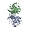
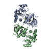
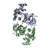
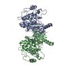
| Entry | Database: PDB / ID: 1uwh | ||||||
|---|---|---|---|---|---|---|---|
| Title | The complex of wild type B-RAF and BAY439006. | ||||||
 Components Components | B-RAF PROTO-ONCOGENE SERINE/THREONINE-PROTEIN KINASE | ||||||
 Keywords Keywords |  TRANSFERASE TRANSFERASE | ||||||
| Function / homology |  Function and homology information Function and homology informationtrehalose metabolism in response to stress / CD4-positive, alpha-beta T cell differentiation / CD4-positive or CD8-positive, alpha-beta T cell lineage commitment / negative regulation of synaptic vesicle exocytosis / head morphogenesis / Signalling to p38 via RIT and RIN / myeloid progenitor cell differentiation / ARMS-mediated activation / SHOC2 M1731 mutant abolishes MRAS complex function / Gain-of-function MRAS complexes activate RAF signaling ...trehalose metabolism in response to stress / CD4-positive, alpha-beta T cell differentiation / CD4-positive or CD8-positive, alpha-beta T cell lineage commitment / negative regulation of synaptic vesicle exocytosis / head morphogenesis / Signalling to p38 via RIT and RIN / myeloid progenitor cell differentiation / ARMS-mediated activation / SHOC2 M1731 mutant abolishes MRAS complex function / Gain-of-function MRAS complexes activate RAF signaling / endothelial cell apoptotic process / negative regulation of fibroblast migration / positive regulation of glucose transmembrane transport / establishment of protein localization to membrane / mitogen-activated protein kinase kinase binding / regulation of T cell differentiation / Negative feedback regulation of MAPK pathway / positive regulation of axonogenesis / Frs2-mediated activation / stress fiber assembly / positive regulation of axon regeneration / face development / synaptic vesicle exocytosis / somatic stem cell population maintenance / MAP kinase kinase activity / thyroid gland development / MAP kinase kinase kinase activity / negative regulation of endothelial cell apoptotic process / positive regulation of substrate adhesion-dependent cell spreading / positive regulation of stress fiber assembly / response to cAMP / ERK1 and ERK2 cascade / cellular response to calcium ion / substrate adhesion-dependent cell spreading / cellular response to nerve growth factor stimulus / thymus development / long-term synaptic potentiation / animal organ morphogenesis / Spry regulation of FGF signaling / RAF activation / Signaling by high-kinase activity BRAF mutants / visual learning / MAP2K and MAPK activation / epidermal growth factor receptor signaling pathway / response to peptide hormone / Negative regulation of MAPK pathway / Signaling by RAF1 mutants / Signaling by moderate kinase activity BRAF mutants / Paradoxical activation of RAF signaling by kinase inactive BRAF / Signaling downstream of RAS mutants / MAPK cascade / Signaling by BRAF and RAF1 fusions / cellular response to xenobiotic stimulus / presynapse / cell body / positive regulation of peptidyl-serine phosphorylation / T cell differentiation in thymus / regulation of cell population proliferation / T cell receptor signaling pathway / scaffold protein binding / negative regulation of neuron apoptotic process / positive regulation of ERK1 and ERK2 cascade / non-specific serine/threonine protein kinase / neuron projection / protein kinase activity / protein phosphorylation / protein serine kinase activity / intracellular membrane-bounded organelle / protein serine/threonine kinase activity / calcium ion binding / protein-containing complex binding / positive regulation of gene expression / negative regulation of apoptotic process / mitochondrion / ATP binding / identical protein binding / nucleus / plasma membrane / cytosolSimilarity search - Function | ||||||
| Biological species |  HOMO SAPIENS (human) HOMO SAPIENS (human) | ||||||
| Method | X-RAY DIFFRACTION / SYNCHROTRON / MOLECULAR REPLACEMENT / Resolution: 2.95 Å | ||||||
 Authors Authors | Barford, D. / Roe, S.M. / Wan, P.T.C. / Cancer Genome Project | ||||||
 Citation Citation | Journal: Cell(Cambridge,Mass.) / Year: 2004 Title: Mechanism of Activation of the Raf-Erk Signaling Pathway by Oncogenic Mutations of B-Raf Authors: Wan, P.T.C. / Garnett, M.J. / Roe, S.M. / Lee, S. / Niculescu-Duvaz, D. / Good, V.M. / Jones, C.M. / Marshall, C.J. / Springer, C.J. / Barford, D. / Marais, R. | ||||||
| History |
|
- Structure visualization
| Structure viewer | Molecule: MolmilJmol/JSmol |
|---|
- Downloads & links
-Download
| PDBx/mmCIF format | 1uwh.cif.gz | 118 KB | Display | PDBx/mmCIF format |
|---|---|---|---|---|
| PDB format | pdb1uwh.ent.gz | 91.8 KB | Display | PDB format |
| PDBx/mmJSON format | 1uwh.json.gz | Tree view | PDBx/mmJSON format | |
| Others |  Other downloads Other downloads |
-Validation report
| Arichive directory | https://data.pdbj.org/pub/pdb/validation_reports/uw/1uwhftp://data.pdbj.org/pub/pdb/validation_reports/uw/1uwh | HTTPS FTP |
|---|
-Related structure data
| Related structure data | 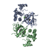 1uwjC  1jpaS 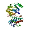 1wfcS  3lckS C: citing same article ( S: Starting model for refinement |
|---|---|
| Similar structure data |

-Links
 PDBj
PDBj


- Assembly
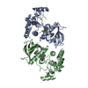
| Deposited unit | 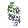
| ||||||||
|---|---|---|---|---|---|---|---|---|---|
| 1 |
| ||||||||
| Unit cell |
|
-Components
| #1: Protein | Mass: 31422.406 Da / Num. of mol.: 2 / Fragment: KINASE DOMAIN, RESIDUES 447-722 Source method: isolated from a genetically manipulated source Source: (gene. exp.) HOMO SAPIENS (human) / Production host:   SPODOPTERA FRUGIPERDA (fall armyworm) SPODOPTERA FRUGIPERDA (fall armyworm)References: UniProt: P15056, EC: 2.7.1.37, non-specific serine/threonine protein kinase#2: Chemical | Sorafenib  Mass: 464.825 Da / Num. of mol.: 2 / Source method: obtained synthetically / Formula: C21H16ClF3N4O3 / Comment: inhibitor*YM Mass: 464.825 Da / Num. of mol.: 2 / Source method: obtained synthetically / Formula: C21H16ClF3N4O3 / Comment: inhibitor*YM#3: Chemical | ChemComp-CL / | Chloride  Mass: 35.453 Da / Num. of mol.: 1 / Source method: obtained synthetically / Formula: Cl Mass: 35.453 Da / Num. of mol.: 1 / Source method: obtained synthetically / Formula: Cl#4: Water | ChemComp-HOH / | Water Mass: 18.015 Da / Num. of mol.: 33 / Source method: isolated from a natural source / Formula: H2O Mass: 18.015 Da / Num. of mol.: 33 / Source method: isolated from a natural source / Formula: H2OCompound details | INVOLVED IN MITOGENIC SIGNAL TRANSDUCTI | |
|---|
-Experimental details
-Experiment
| Experiment | Method: X-RAY DIFFRACTION / Number of used crystals: 1 |
|---|
- Sample preparation
| Crystal | Density Matthews: 3.2 Å3/Da / Density % sol: 62 % | ||||||||||||||||||
|---|---|---|---|---|---|---|---|---|---|---|---|---|---|---|---|---|---|---|---|
| Crystal grow | pH: 8.5 / Details: pH 8.50 | ||||||||||||||||||
| Crystal grow | *PLUS Temperature: 20 ℃ / pH: 8.5 / Method: batch method | ||||||||||||||||||
| Components of the solutions | *PLUS
|
-Data collection
| Diffraction | Mean temperature: 100 K |
|---|---|
| Diffraction source | Source: SYNCHROTRON / Site: ESRF  / Beamline: ID13 / Wavelength: 0.9755 / Beamline: ID13 / Wavelength: 0.9755 |
| Detector | Type: MARRESEARCH / Detector: CCD / Date: Mar 15, 2003 |
| Radiation | Protocol: SINGLE WAVELENGTH / Monochromatic (M) / Laue (L): M / Scattering type: x-ray |
| Radiation wavelength | Wavelength: 0.9755 Å / Relative weight: 1 |
| Reflection | Resolution: 2.95→30 Å / Num. obs: 18727 / % possible obs: 98 % / Redundancy: 3.5 % / Biso Wilson estimate: 106.7 Å2 / Rmerge(I) obs: 0.123 / Net I/σ(I): 8.8 |
| Reflection shell | Resolution: 2.95→3.07 Å / Redundancy: 2.6 % / Rmerge(I) obs: 0.351 / Mean I/σ(I) obs: 2.1 / % possible all: 98 |
| Reflection | *PLUS Highest resolution: 2.95 Å / Lowest resolution: 30 Å / Redundancy: 3.5 % / Num. measured all: 66126 / Rmerge(I) obs: 0.123 |
| Reflection shell | *PLUS % possible obs: 98 % / Redundancy: 2.6 % / Num. unique obs: 1968 / Num. measured obs: 5030 / Rmerge(I) obs: 0.351 / Mean I/σ(I) obs: 1.8 |
- Processing
| Software |
| ||||||||||||||||||||||||||||||||||||||||||||||||||||||||||||||||||||||||||||||||
|---|---|---|---|---|---|---|---|---|---|---|---|---|---|---|---|---|---|---|---|---|---|---|---|---|---|---|---|---|---|---|---|---|---|---|---|---|---|---|---|---|---|---|---|---|---|---|---|---|---|---|---|---|---|---|---|---|---|---|---|---|---|---|---|---|---|---|---|---|---|---|---|---|---|---|---|---|---|---|---|---|---|
| Refinement | Method to determine structure: MOLECULAR REPLACEMENT Starting model: PDB ENTRY 1WFC, 3LCK, 1JPA Resolution: 2.95→29.91 Å / Rfactor Rfree error: 0.009 / Data cutoff high absF: 1710100.43 / Isotropic thermal model: RESTRAINED / Cross valid method: THROUGHOUT / σ(F): 0 Details: RESIDUES AT THE N AND C-TERMINI AND PART OF THE ACTIVATION LOOP (600-611) ARE DISORDERED AND NOT SEEN.
| ||||||||||||||||||||||||||||||||||||||||||||||||||||||||||||||||||||||||||||||||
| Solvent computation | Solvent model: FLAT MODEL / Bsol: 24.6495 Å2 / ksol: 0.329116 e/Å3 | ||||||||||||||||||||||||||||||||||||||||||||||||||||||||||||||||||||||||||||||||
| Displacement parameters | Biso mean: 59 Å2
| ||||||||||||||||||||||||||||||||||||||||||||||||||||||||||||||||||||||||||||||||
| Refine analyze |
| ||||||||||||||||||||||||||||||||||||||||||||||||||||||||||||||||||||||||||||||||
| Refinement step | Cycle: LAST / Resolution: 2.95→29.91 Å
| ||||||||||||||||||||||||||||||||||||||||||||||||||||||||||||||||||||||||||||||||
| Refine LS restraints |
| ||||||||||||||||||||||||||||||||||||||||||||||||||||||||||||||||||||||||||||||||
| LS refinement shell | Resolution: 2.95→3.13 Å / Rfactor Rfree error: 0.028 / Total num. of bins used: 6
| ||||||||||||||||||||||||||||||||||||||||||||||||||||||||||||||||||||||||||||||||
| Xplor file |
| ||||||||||||||||||||||||||||||||||||||||||||||||||||||||||||||||||||||||||||||||
| Refinement | *PLUS Rfactor Rfree: 0.251 / Rfactor Rwork: 0.217 | ||||||||||||||||||||||||||||||||||||||||||||||||||||||||||||||||||||||||||||||||
| Solvent computation | *PLUS | ||||||||||||||||||||||||||||||||||||||||||||||||||||||||||||||||||||||||||||||||
| Displacement parameters | *PLUS | ||||||||||||||||||||||||||||||||||||||||||||||||||||||||||||||||||||||||||||||||
| Refine LS restraints | *PLUS
|
