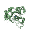
[English] 日本語
Yorodumi
- PDB-1unf: The crystal structure of the eukaryotic FeSOD from Vigna unguicul... -

+
Open data


ID or keywords:

Loading...

-
Basic information

Entry
Database: PDB / ID: 1unf
TitleThe crystal structure of the eukaryotic FeSOD from Vigna unguiculata suggests a new enzymatic mechanism
ComponentsIRON SUPEROXIDE DISMUTASE
KeywordsOXIDOREDUCTASE / EUKARYOTIC / SUPEROXIDE DISMUTASE / METALLOPROTEIN
Function / homology
Function and homology information


superoxide dismutase / superoxide dismutase activity / metal ion binding
Similarity search - Function
Manganese/iron superoxide dismutase, binding site / Manganese and iron superoxide dismutases signature. / Manganese/iron superoxide dismutase / Manganese/iron superoxide dismutase, N-terminal / Iron/manganese superoxide dismutases, alpha-hairpin domain / Manganese/iron superoxide dismutase, C-terminal / Manganese/iron superoxide dismutase, C-terminal domain superfamily / Iron/manganese superoxide dismutases, C-terminal domain / Manganese/iron superoxide dismutase, N-terminal domain superfamily
Similarity search - Domain/homology
: / Superoxide dismutase
Similarity search - Component
Biological speciesVIGNA UNGUICULATA (cowpea)
MethodX-RAY DIFFRACTION / SYNCHROTRON / MOLECULAR REPLACEMENT / Resolution: 1.97 Å
AuthorsMunoz, I.G. / Moran, J.F. / Becana, M. / Montoya, G.
CitationJournal: Protein Sci. / Year: 2005
Title: The Crystal Structure of an Eukaryotic Iron Superoxide Dismutase Suggests Intersubunit Cooperation During Catalysis
Authors: Munoz, I.G. / Moran, J.F. / Becana, M. / Montoya, G.
History
DepositionSep 10, 2003Deposition site: PDBE / Processing site: PDBE
Revision 1.0Oct 27, 2004Provider: repository / Type: Initial release
Revision 1.1May 8, 2011Group: Version format compliance
Revision 1.2Jul 13, 2011Group: Version format compliance
Revision 1.3Dec 13, 2023Group: Data collection / Database references ...Data collection / Database references / Derived calculations / Refinement description
Category: chem_comp_atom / chem_comp_bond ...chem_comp_atom / chem_comp_bond / database_2 / pdbx_initial_refinement_model / pdbx_struct_conn_angle / struct_conn
Item: _database_2.pdbx_DOI / _database_2.pdbx_database_accession ..._database_2.pdbx_DOI / _database_2.pdbx_database_accession / _pdbx_struct_conn_angle.ptnr1_auth_comp_id / _pdbx_struct_conn_angle.ptnr1_auth_seq_id / _pdbx_struct_conn_angle.ptnr1_label_atom_id / _pdbx_struct_conn_angle.ptnr1_label_comp_id / _pdbx_struct_conn_angle.ptnr1_label_seq_id / _pdbx_struct_conn_angle.ptnr3_auth_comp_id / _pdbx_struct_conn_angle.ptnr3_auth_seq_id / _pdbx_struct_conn_angle.ptnr3_label_atom_id / _pdbx_struct_conn_angle.ptnr3_label_comp_id / _pdbx_struct_conn_angle.ptnr3_label_seq_id / _pdbx_struct_conn_angle.value / _struct_conn.pdbx_dist_value / _struct_conn.ptnr1_auth_comp_id / _struct_conn.ptnr1_auth_seq_id / _struct_conn.ptnr1_label_asym_id / _struct_conn.ptnr1_label_atom_id / _struct_conn.ptnr1_label_comp_id / _struct_conn.ptnr1_label_seq_id / _struct_conn.ptnr2_auth_comp_id / _struct_conn.ptnr2_auth_seq_id / _struct_conn.ptnr2_label_asym_id / _struct_conn.ptnr2_label_atom_id / _struct_conn.ptnr2_label_comp_id / _struct_conn.ptnr2_label_seq_id

-
Structure visualization

Structure viewerMolecule:
MolmilJmol/JSmol

Downloads & links

-
Assembly

Deposited unit
X: IRON SUPEROXIDE DISMUTASE
hetero molecules


Theoretical massNumber of molelcules
Total (without water)26,7952
Polymers26,7391
Non-polymers561
Water3,315184
1


  • Idetical with deposited unit
  • defined by author&software
TypeNameSymmetry operationNumber
identity operation1_555x,y,z1
MethodPQS
Unit cell
Length a, b, c (Å)81.985, 48.163, 63.671
Angle α, β, γ (deg.)90.00, 119.76, 90.00
Int Tables number5
Space group name H-MC121
Components on special symmetry positions
IDModelComponents
11X-2135-

HOH

-
Components

#1: Protein IRON SUPEROXIDE DISMUTASE / / SUPEROXIDE DISMUTASE [MN/FE]


Mass: 26739.188 Da / Num. of mol.: 1 / Source method: isolated from a natural source / Source: (natural) VIGNA UNGUICULATA (cowpea) / Cellular location: CYTOSOL / Strain: VUFESOD / Tissue: LEGUME ROOT NODULES / References: UniProt: Q9M7R2, superoxide dismutase
#2: Chemical ChemComp-FE / FE (III) ION / Iron


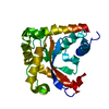
Mass: 55.845 Da / Num. of mol.: 1 / Source method: obtained synthetically / Formula: Fe
#3: Water ChemComp-HOH / water / Water


Mass: 18.015 Da / Num. of mol.: 184 / Source method: isolated from a natural source / Formula: H2O

-
Experimental details

-
Experiment

ExperimentMethod: X-RAY DIFFRACTION / Number of used crystals: 1

-
Sample preparation

CrystalDensity Matthews: 2.23 Å3/Da / Density % sol: 44.46 %
Crystal growpH: 5
Details: 200 MM AMMONIUM SULFATE, 100 MM SODIUM ACETATE PH 5.0, 25%(W/V) POLYETHYLENE GLYCOL 4000

-
Data collection

DiffractionMean temperature: 100 K
Diffraction sourceSource: SYNCHROTRON / Site: ESRF / Beamline: BM14 / Wavelength: 1.033
DetectorType: MARRESEARCH / Detector: CCD / Date: Nov 15, 2002
RadiationProtocol: SINGLE WAVELENGTH / Monochromatic (M) / Laue (L): M / Scattering type: x-ray
Radiation wavelengthWavelength: 1.033 Å / Relative weight: 1
ReflectionResolution: 1.8→30 Å / Num. obs: 20443 / % possible obs: 99 % / Observed criterion σ(I): 2 / Redundancy: 3.3 % / Rmerge(I) obs: 0.1 / Net I/σ(I): 6.2
Reflection shellResolution: 1.8→1.9 Å / Redundancy: 3.1 % / Rmerge(I) obs: 0.77 / Mean I/σ(I) obs: 0.9 / % possible all: 99

-
Processing

Software
NameVersionClassification
REFMAC5.2.0005refinement
MOSFLMdata reduction
SCALAdata scaling
EPMRphasing
RefinementMethod to determine structure: MOLECULAR REPLACEMENT
Starting model: PDB ENTRY 1DT0
Resolution: 1.97→28.77 Å / Cor.coef. Fo:Fc: 0.961 / Cor.coef. Fo:Fc free: 0.946 / SU B: 6.356 / SU ML: 0.084 / Cross valid method: THROUGHOUT / ESU R Free: 0.139
Stereochemistry target values: MAXIMUM LIKELIHOOD WITH PHASES
Details: HYDROGENS HAVE BEEN ADDED IN THE RIDING POSITIONS
RfactorNum. reflection% reflectionSelection details
Rfree0.192 772 5 %RANDOM
Rwork0.148 ---
obs0.15 14661 99.6 %-
Solvent computationIon probe radii: 0.8 Å / Shrinkage radii: 0.8 Å / VDW probe radii: 1.2 Å / Solvent model: BABINET MODEL WITH MASK
Displacement parametersBiso mean: 22.65 Å2
Baniso -1Baniso -2Baniso -3
1--0.03 Å20 Å20 Å2
2--0.02 Å20 Å2
3---0.01 Å2
Refinement stepCycle: LAST / Resolution: 1.97→28.77 Å
ProteinNucleic acidLigandSolventTotal
Num. atoms1724 0 1 184 1909
Refine LS restraints
Refine-IDTypeDev idealDev ideal targetNumber
X-RAY DIFFRACTIONr_bond_refined_d0.0140.0221775
X-RAY DIFFRACTIONr_bond_other_d
X-RAY DIFFRACTIONr_angle_refined_deg1.5851.9432409
X-RAY DIFFRACTIONr_angle_other_deg
X-RAY DIFFRACTIONr_dihedral_angle_1_deg7.7645211
X-RAY DIFFRACTIONr_dihedral_angle_2_deg33.37524.82485
X-RAY DIFFRACTIONr_dihedral_angle_3_deg16.26215289
X-RAY DIFFRACTIONr_dihedral_angle_4_deg12.073155
X-RAY DIFFRACTIONr_chiral_restr0.190.2249
X-RAY DIFFRACTIONr_gen_planes_refined0.0060.021371
X-RAY DIFFRACTIONr_gen_planes_other
X-RAY DIFFRACTIONr_nbd_refined0.2280.3940
X-RAY DIFFRACTIONr_nbd_other
X-RAY DIFFRACTIONr_nbtor_refined0.3230.51227
X-RAY DIFFRACTIONr_nbtor_other
X-RAY DIFFRACTIONr_xyhbond_nbd_refined0.2050.5242
X-RAY DIFFRACTIONr_xyhbond_nbd_other
X-RAY DIFFRACTIONr_metal_ion_refined0.0760.51
X-RAY DIFFRACTIONr_metal_ion_other
X-RAY DIFFRACTIONr_symmetry_vdw_refined0.1920.331
X-RAY DIFFRACTIONr_symmetry_vdw_other
X-RAY DIFFRACTIONr_symmetry_hbond_refined0.3620.529
X-RAY DIFFRACTIONr_symmetry_hbond_other
X-RAY DIFFRACTIONr_symmetry_metal_ion_refined
X-RAY DIFFRACTIONr_symmetry_metal_ion_other
X-RAY DIFFRACTIONr_mcbond_it2.28121091
X-RAY DIFFRACTIONr_mcbond_other
X-RAY DIFFRACTIONr_mcangle_it3.3831706
X-RAY DIFFRACTIONr_mcangle_other
X-RAY DIFFRACTIONr_scbond_it2.4342807
X-RAY DIFFRACTIONr_scbond_other
X-RAY DIFFRACTIONr_scangle_it3.4413703
X-RAY DIFFRACTIONr_scangle_other
X-RAY DIFFRACTIONr_long_range_B_refined
X-RAY DIFFRACTIONr_long_range_B_other
X-RAY DIFFRACTIONr_rigid_bond_restr
X-RAY DIFFRACTIONr_sphericity_free
X-RAY DIFFRACTIONr_sphericity_bonded
LS refinement shellResolution: 1.97→2.02 Å / Total num. of bins used: 20
RfactorNum. reflection% reflection
Rfree0.215 57 -
Rwork0.146 1024 -
obs--96.17 %

+
About Yorodumi

-
News

-
Feb 9, 2022. New format data for meta-information of EMDB entries

New format data for meta-information of EMDB entries

  • Version 3 of the EMDB header file is now the official format.
  • The previous official version 1.9 will be removed from the archive.

Related info.:EMDB header

External links:wwPDB to switch to version 3 of the EMDB data model

-
Aug 12, 2020. Covid-19 info

Covid-19 info

URL: https://pdbj.org/emnavi/covid19.php

New page: Covid-19 featured information page in EM Navigator.

Related info.:Covid-19 info / Mar 5, 2020. Novel coronavirus structure data

+
Mar 5, 2020. Novel coronavirus structure data

Novel coronavirus structure data

Related info.:Yorodumi Speices / Aug 12, 2020. Covid-19 info

External links:COVID-19 featured content - PDBj / Molecule of the Month (242):Coronavirus Proteases

+
Jan 31, 2019. EMDB accession codes are about to change! (news from PDBe EMDB page)

EMDB accession codes are about to change! (news from PDBe EMDB page)

  • The allocation of 4 digits for EMDB accession codes will soon come to an end. Whilst these codes will remain in use, new EMDB accession codes will include an additional digit and will expand incrementally as the available range of codes is exhausted. The current 4-digit format prefixed with “EMD-” (i.e. EMD-XXXX) will advance to a 5-digit format (i.e. EMD-XXXXX), and so on. It is currently estimated that the 4-digit codes will be depleted around Spring 2019, at which point the 5-digit format will come into force.
  • The EM Navigator/Yorodumi systems omit the EMD- prefix.

Related info.:Q: What is EMD? / ID/Accession-code notation in Yorodumi/EM Navigator

External links:EMDB Accession Codes are Changing Soon! / Contact to PDBj

+
Jul 12, 2017. Major update of PDB

Major update of PDB

  • wwPDB released updated PDB data conforming to the new PDBx/mmCIF dictionary.
  • This is a major update changing the version number from 4 to 5, and with Remediation, in which all the entries are updated.
  • In this update, many items about electron microscopy experimental information are reorganized (e.g. em_software).
  • Now, EM Navigator and Yorodumi are based on the updated data.

External links:wwPDB Remediation / Enriched Model Files Conforming to OneDep Data Standards Now Available in the PDB FTP Archive

-
Yorodumi

Thousand views of thousand structures

  • Yorodumi is a browser for structure data from EMDB, PDB, SASBDB, etc.
  • This page is also the successor to EM Navigator detail page, and also detail information page/front-end page for Omokage search.
  • The word "yorodu" (or yorozu) is an old Japanese word meaning "ten thousand". "mi" (miru) is to see.

Related info.:EMDB / PDB / SASBDB / Comparison of 3 databanks / Yorodumi Search / Aug 31, 2016. New EM Navigator & Yorodumi / Yorodumi Papers / Jmol/JSmol / Function and homology information / Changes in new EM Navigator and Yorodumi

Read more