 Movie
Movie Controller


+ Open data

- Basic information
| Entry | Database: PDB / ID: 1umy | ||||||
|---|---|---|---|---|---|---|---|
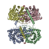
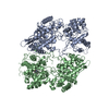
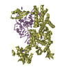
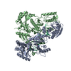
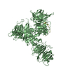
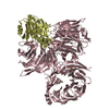
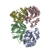
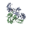
| Title | BHMT from rat liver | ||||||
 Components Components | Betaine--homocysteine S-methyltransferase 1 | ||||||
 Keywords Keywords | TRANSFERASE / METHIONINE SYNTHESIS / HOMOCYSTEINE METABOLISM / BETAINE / METHYLTRANSFERASE / ZINC | ||||||
| Function / homology |  Function and homology information Function and homology informationCholine catabolism / betaine-homocysteine S-methyltransferase / amino-acid betaine metabolic process / betaine-homocysteine S-methyltransferase activity / L-methionine salvage / Sulfur amino acid metabolism / amino-acid betaine catabolic process / response to alcohol / methyltransferase activity / methylation ...Choline catabolism / betaine-homocysteine S-methyltransferase / amino-acid betaine metabolic process / betaine-homocysteine S-methyltransferase activity / L-methionine salvage / Sulfur amino acid metabolism / amino-acid betaine catabolic process / response to alcohol / methyltransferase activity / methylation / protein-containing complex binding / protein-containing complex / extracellular space / extracellular exosome / zinc ion binding / identical protein binding / nucleus / cytosol Similarity search - Function | ||||||
| Biological species |  | ||||||
| Method |  X-RAY DIFFRACTION / SYNCHROTRON / MOLECULAR REPLACEMENT / Resolution: 2.5 Å X-RAY DIFFRACTION / SYNCHROTRON / MOLECULAR REPLACEMENT / Resolution: 2.5 Å | ||||||
 Authors Authors | Gonzalez, B. / Pajares, M.A. / Sanz-Aparicio, J. | ||||||
 Citation Citation | Journal: J. Mol. Biol. / Year: 2004 Title: Crystal structure of rat liver betaine homocysteine s-methyltransferase reveals new oligomerization features and conformational changes upon substrate binding. Authors: Gonzalez, B. / Pajares, M.A. / Martinez-Ripoll, M. / Blundell, T.L. / Sanz-Aparicio, J. #1: Journal: Biochem.J. / Year: 2003 Title: Active-Site Mutagenesis Study of Rat Liver Betaine Homocysteine S-Methyltransferase Authors: Gonzalez, B. / Garrido, F. / Gasset, M. / Sanz-Aparicio, J. / Pajares, M.A. #2: Journal: Acta Crystallogr.,Sect.D / Year: 2002 Title: Crystallization and Preliminary X-Ray Study of Recombinant Betaine-Homocysteine S-Methyltransferase from Rat Liver Authors: Gonzalez, B. / Pajares, M.A. / Too, P. / Garrido, F. / Blundell, T.L. / Sanz-Aparicio, J. | ||||||
| History |
|
- Structure visualization
| Structure viewer | Molecule: MolmilJmol/JSmol |
|---|
- Downloads & links
-Download
| PDBx/mmCIF format | 1umy.cif.gz | 309.1 KB | Display | PDBx/mmCIF format |
|---|---|---|---|---|
| PDB format | pdb1umy.ent.gz | 249 KB | Display | PDB format |
| PDBx/mmJSON format | 1umy.json.gz | Tree view | PDBx/mmJSON format | |
| Others |  Other downloads Other downloads |
-Validation report
| Arichive directory | https://data.pdbj.org/pub/pdb/validation_reports/um/1umyftp://data.pdbj.org/pub/pdb/validation_reports/um/1umy | HTTPS FTP |
|---|
-Related structure data
| Related structure data | 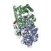 1lt8S S: Starting model for refinement |
|---|---|
| Similar structure data |

-Links
 PDBj
PDBj


- Assembly
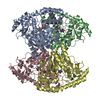
| Deposited unit | 
| ||||||||||||||||
|---|---|---|---|---|---|---|---|---|---|---|---|---|---|---|---|---|---|
| 1 |
| ||||||||||||||||
| Unit cell |
| ||||||||||||||||
| Noncrystallographic symmetry (NCS) | NCS oper:
|
-Components
| #1: Protein | Mass: 45055.410 Da / Num. of mol.: 4 Source method: isolated from a genetically manipulated source Details: ZN BOUND FORM / Source: (gene. exp.)  References: UniProt: O09171, betaine-homocysteine S-methyltransferase #2: Chemical | ChemComp-ZN /   Mass: 65.409 Da / Num. of mol.: 4 / Source method: obtained synthetically / Formula: Zn Mass: 65.409 Da / Num. of mol.: 4 / Source method: obtained synthetically / Formula: Zn#3: Chemical |   Mass: 78.133 Da / Num. of mol.: 2 / Source method: obtained synthetically / Formula: C2H6OS Mass: 78.133 Da / Num. of mol.: 2 / Source method: obtained synthetically / Formula: C2H6OS#4: Water | ChemComp-HOH / |  Mass: 18.015 Da / Num. of mol.: 538 / Source method: isolated from a natural source / Formula: H2O Mass: 18.015 Da / Num. of mol.: 538 / Source method: isolated from a natural source / Formula: H2OCompound details | INVOLVED IN THE REGULATION OF HOMOCYSTEINE METABOLISM. CONVERTS HOMOCYSTEINE AND BETAINE TO ...INVOLVED IN THE REGULATION | Has protein modification | Y | |
|---|
-Experimental details
-Experiment
| Experiment | Method: X-RAY DIFFRACTION / Number of used crystals: 1 |
|---|
- Sample preparation
| Crystal | Density Matthews: 2.2 Å3/Da / Density % sol: 44 % |
|---|---|
| Crystal grow | pH: 8.5 / Details: 16% PEG 1500, PH=8.5, pH 8.50 |
-Data collection
| Diffraction | Mean temperature: 100 K |
|---|---|
| Diffraction source | Source: SYNCHROTRON / Site: ESRF  / Beamline: BM14 / Wavelength: 1.004 / Beamline: BM14 / Wavelength: 1.004 |
| Detector | Detector: CCD / Date: Apr 15, 2001 |
| Radiation | Protocol: SINGLE WAVELENGTH / Monochromatic (M) / Laue (L): M / Scattering type: x-ray |
| Radiation wavelength | Wavelength: 1.004 Å / Relative weight: 1 |
| Reflection | Resolution: 2.5→49 Å / Num. obs: 58532 / % possible obs: 99.7 % / Observed criterion σ(I): 4 / Redundancy: 4 % / Biso Wilson estimate: 45.8 Å2 / Rmerge(I) obs: 0.088 / Net I/σ(I): 6.5 |
| Reflection shell | Resolution: 2.5→2.58 Å / Redundancy: 4 % / Rmerge(I) obs: 0.46 / Mean I/σ(I) obs: 1.5 / % possible all: 99.1 |
- Processing
| Software |
| ||||||||||||||||||||||||||||||||||||||||||||||||||||||||||||
|---|---|---|---|---|---|---|---|---|---|---|---|---|---|---|---|---|---|---|---|---|---|---|---|---|---|---|---|---|---|---|---|---|---|---|---|---|---|---|---|---|---|---|---|---|---|---|---|---|---|---|---|---|---|---|---|---|---|---|---|---|---|
| Refinement | Method to determine structure: MOLECULAR REPLACEMENT Starting model: PDB ENTRY 1LT8 Resolution: 2.5→48.4 Å / Rfactor Rfree error: 0.004 / Data cutoff high absF: 10000000 / Cross valid method: THROUGHOUT / σ(F): 0
| ||||||||||||||||||||||||||||||||||||||||||||||||||||||||||||
| Solvent computation | Solvent model: FLAT / Bsol: 61.82 Å2 / ksol: 0.356894 e/Å3 | ||||||||||||||||||||||||||||||||||||||||||||||||||||||||||||
| Displacement parameters | Biso mean: 50.9 Å2 | ||||||||||||||||||||||||||||||||||||||||||||||||||||||||||||
| Refine analyze |
| ||||||||||||||||||||||||||||||||||||||||||||||||||||||||||||
| Refinement step | Cycle: LAST / Resolution: 2.5→48.4 Å
| ||||||||||||||||||||||||||||||||||||||||||||||||||||||||||||
| Refine LS restraints |
| ||||||||||||||||||||||||||||||||||||||||||||||||||||||||||||
| LS refinement shell | Resolution: 2.5→2.66 Å / Rfactor Rfree error: 0.013 / Total num. of bins used: 10
| ||||||||||||||||||||||||||||||||||||||||||||||||||||||||||||
| Xplor file |
|
