 Movie
Movie Controller

[English] 日本語
 Yorodumi
- PDB-1umd: branched-chain 2-oxo acid dehydrogenase (E1) from Thermus thermop... -
+ Open data

- Basic information
| Entry | Database: PDB / ID: 1umd | |||||||||
|---|---|---|---|---|---|---|---|---|---|---|
| Title | branched-chain 2-oxo acid dehydrogenase (E1) from Thermus thermophilus HB8 with 4-methyl-2-oxopentanoate as an intermediate | |||||||||
 Components Components | (2-oxo acid dehydrogenase ...) x 2 | |||||||||
 Keywords Keywords | OXIDOREDUCTASE / alpha(2)beta(2) tetramer / structural genomics / RIKEN Structural Genomics/Proteomics Initiative / RSGI | |||||||||
| Function / homology |  Function and homology information Function and homology information3-methyl-2-oxobutanoate dehydrogenase (2-methylpropanoyl-transferring) / branched-chain 2-oxo acid dehydrogenase activity / branched-chain amino acid catabolic process / metal ion binding Similarity search - Function | |||||||||
| Biological species |   Thermus thermophilus (bacteria) Thermus thermophilus (bacteria) | |||||||||
| Method |  X-RAY DIFFRACTION / SYNCHROTRON / MOLECULAR REPLACEMENT / Resolution: 1.9 Å X-RAY DIFFRACTION / SYNCHROTRON / MOLECULAR REPLACEMENT / Resolution: 1.9 Å | |||||||||
 Authors Authors | Nakai, T. / Nakagawa, N. / Maoka, N. / Masui, R. / Kuramitsu, S. / Kamiya, N. / RIKEN Structural Genomics/Proteomics Initiative (RSGI) | |||||||||
 Citation Citation | Journal: J.Mol.Biol. / Year: 2004 Title: Ligand-induced Conformational Changes and a Reaction Intermediate in Branched-chain 2-Oxo Acid Dehydrogenase (E1) from Thermus thermophilus HB8, as Revealed by X-ray Crystallography Authors: Nakai, T. / Nakagawa, N. / Maoka, N. / Masui, R. / Kuramitsu, S. / Kamiya, N. | |||||||||
| History |
|
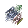
- Structure visualization
| Structure viewer | Molecule: MolmilJmol/JSmol |
|---|
- Downloads & links
-Download
| PDBx/mmCIF format | 1umd.cif.gz | 306.6 KB | Display | PDBx/mmCIF format |
|---|---|---|---|---|
| PDB format | pdb1umd.ent.gz | 242.1 KB | Display | PDB format |
| PDBx/mmJSON format | 1umd.json.gz | Tree view | PDBx/mmJSON format | |
| Others |  Other downloads Other downloads |
-Validation report
| Arichive directory | https://data.pdbj.org/pub/pdb/validation_reports/um/1umdftp://data.pdbj.org/pub/pdb/validation_reports/um/1umd | HTTPS FTP |
|---|
-Related structure data
| Related structure data | 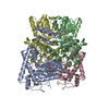 1um9C  1umbC 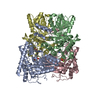 1umcC 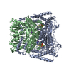 1dtwS C: citing same article ( S: Starting model for refinement |
|---|---|
| Similar structure data | |
| Other databases |


-Links
 PDBj
PDBj


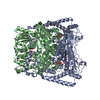
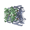
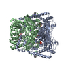
- Assembly
| Deposited unit | 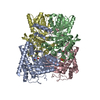
| ||||||||
|---|---|---|---|---|---|---|---|---|---|
| 1 |
| ||||||||
| Unit cell |
| ||||||||
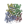
| Details | The biological assembly is an alpha(2)beta(2) tetramer in the asymmetric unit. |
-Components
-2-oxo acid dehydrogenase ... , 2 types, 4 molecules ACBD
| #1: Protein | Mass: 41508.023 Da / Num. of mol.: 2 Source method: isolated from a genetically manipulated source Source: (gene. exp.) Thermus thermophilus (bacteria) / Plasmid: pACYC184 / Production host: References: UniProt: Q5SLR4, 3-methyl-2-oxobutanoate dehydrogenase (2-methylpropanoyl-transferring) #2: Protein | Mass: 35180.582 Da / Num. of mol.: 2 Source method: isolated from a genetically manipulated source Source: (gene. exp.) Thermus thermophilus (bacteria) / Plasmid: pET11A / Production host: References: UniProt: Q5SLR3, 3-methyl-2-oxobutanoate dehydrogenase (2-methylpropanoyl-transferring) |
|---|
-Non-polymers , 4 types, 1340 molecules 

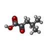

| #3: Chemical |  Mass: 24.305 Da / Num. of mol.: 2 / Source method: obtained synthetically / Formula: Mg Mass: 24.305 Da / Num. of mol.: 2 / Source method: obtained synthetically / Formula: Mg#4: Chemical |  Mass: 425.314 Da / Num. of mol.: 2 / Source method: obtained synthetically / Formula: C12H19N4O7P2S Mass: 425.314 Da / Num. of mol.: 2 / Source method: obtained synthetically / Formula: C12H19N4O7P2S#5: Chemical |  Mass: 130.142 Da / Num. of mol.: 2 / Source method: obtained synthetically / Formula: C6H10O3 Mass: 130.142 Da / Num. of mol.: 2 / Source method: obtained synthetically / Formula: C6H10O3#6: Water | ChemComp-HOH / | Mass: 18.015 Da / Num. of mol.: 1334 / Source method: isolated from a natural source / Formula: H2O |
|---|
-Experimental details
-Experiment
| Experiment | Method: X-RAY DIFFRACTION / Number of used crystals: 1 |
|---|
- Sample preparation
| Crystal | Density Matthews: 3.19 Å3/Da / Density % sol: 61.17 % |
|---|---|
| Crystal grow | Temperature: 291 K / Method: vapor diffusion, hanging drop / pH: 5.6 Details: lithium sulfate, sodium citrate, pH 5.6, VAPOR DIFFUSION, HANGING DROP, temperature 291K |
-Data collection
| Diffraction | Mean temperature: 100 K |
|---|---|
| Diffraction source | Source: SYNCHROTRON / Site: SPring-8  / Beamline: BL45XU / Wavelength: 1.0718 Å / Beamline: BL45XU / Wavelength: 1.0718 Å |
| Detector | Type: RIGAKU RAXIS V / Detector: IMAGE PLATE / Date: Jul 7, 2002 |
| Radiation | Monochromator: Si(111) / Protocol: SINGLE WAVELENGTH / Monochromatic (M) / Laue (L): M / Scattering type: x-ray |
| Radiation wavelength | Wavelength: 1.0718 Å / Relative weight: 1 |
| Reflection | Resolution: 1.9→50 Å / Num. all: 167525 / Num. obs: 167525 / % possible obs: 99.2 % / Observed criterion σ(I): 0 / Redundancy: 4.7 % / Biso Wilson estimate: 12.1 Å2 / Rmerge(I) obs: 0.101 / Net I/σ(I): 8.7 |
| Reflection shell | Resolution: 1.9→1.97 Å / Redundancy: 4 % / Rmerge(I) obs: 0.313 / Mean I/σ(I) obs: 1.8 / Num. unique all: 16506 / % possible all: 98.6 |
- Processing
| Software |
| ||||||||||||||||||||||||||||||||||||||||||||||||||||||||||||
|---|---|---|---|---|---|---|---|---|---|---|---|---|---|---|---|---|---|---|---|---|---|---|---|---|---|---|---|---|---|---|---|---|---|---|---|---|---|---|---|---|---|---|---|---|---|---|---|---|---|---|---|---|---|---|---|---|---|---|---|---|---|
| Refinement | Method to determine structure: MOLECULAR REPLACEMENT Starting model: PDB ENTRY 1DTW Resolution: 1.9→19.99 Å / Rfactor Rfree error: 0.002 / Data cutoff high absF: 4618879.32 / Data cutoff low absF: 0 / Isotropic thermal model: RESTRAINED / Cross valid method: THROUGHOUT / σ(F): 0 / Stereochemistry target values: Engh & Huber
| ||||||||||||||||||||||||||||||||||||||||||||||||||||||||||||
| Solvent computation | Solvent model: FLAT MODEL / Bsol: 55.4141 Å2 / ksol: 0.371797 e/Å3 | ||||||||||||||||||||||||||||||||||||||||||||||||||||||||||||
| Displacement parameters | Biso mean: 19.8 Å2
| ||||||||||||||||||||||||||||||||||||||||||||||||||||||||||||
| Refine analyze |
| ||||||||||||||||||||||||||||||||||||||||||||||||||||||||||||
| Refinement step | Cycle: LAST / Resolution: 1.9→19.99 Å
| ||||||||||||||||||||||||||||||||||||||||||||||||||||||||||||
| Refine LS restraints |
| ||||||||||||||||||||||||||||||||||||||||||||||||||||||||||||
| LS refinement shell | Resolution: 1.9→2.02 Å / Rfactor Rfree error: 0.007 / Total num. of bins used: 6
| ||||||||||||||||||||||||||||||||||||||||||||||||||||||||||||
| Xplor file |
|
