 Movie
Movie Controller

[English] 日本語
 Yorodumi
- PDB-1ucc: Crystal structure of the Ribonuclease MC1 from bitter gourd seeds... -
+ Open data

- Basic information
| Entry | Database: PDB / ID: 1ucc | ||||||
|---|---|---|---|---|---|---|---|
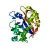
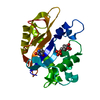
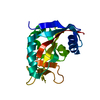
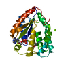
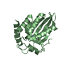
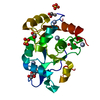
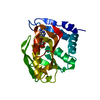
| Title | Crystal structure of the Ribonuclease MC1 from bitter gourd seeds complexed with 3'-UMP. | ||||||
 Components Components | Ribonuclease MC | ||||||
 Keywords Keywords |  HYDROLASE / alpha plus beta HYDROLASE / alpha plus beta | ||||||
| Function / homology |  Function and homology informationribonuclease T2 / ribonuclease T2 activity / organic substance metabolic process / RNA binding Function and homology informationribonuclease T2 / ribonuclease T2 activity / organic substance metabolic process / RNA bindingSimilarity search - Function | ||||||
| Biological species |  Momordica charantia (bitter melon) Momordica charantia (bitter melon) | ||||||
| Method | X-RAY DIFFRACTION / SYNCHROTRON / MOLECULAR REPLACEMENT / Resolution: 1.77 Å | ||||||
 Authors Authors | Suzuki, A. / Yao, M. / Tanaka, I. / Numata, T. / Kikukawa, S. / Yamasaki, N. / Kimura, M. | ||||||
 Citation Citation | Journal: Biochem.Biophys.Res.Commun. / Year: 2000 Title: Crystal structures of the ribonuclease MC1 from bitter gourd seeds, complexed with 2'-UMP or 3'-UMP, reveal structural basis for uridine specificity Authors: Suzuki, A. / Yao, M. / Tanaka, I. / Numata, T. / Kikukawa, S. / Yamasaki, N. / Kimura, M. | ||||||
| History |
|
- Structure visualization
| Structure viewer | Molecule: MolmilJmol/JSmol |
|---|
- Downloads & links
-Download
| PDBx/mmCIF format | 1ucc.cif.gz | 52.5 KB | Display | PDBx/mmCIF format |
|---|---|---|---|---|
| PDB format | pdb1ucc.ent.gz | 36.6 KB | Display | PDB format |
| PDBx/mmJSON format | 1ucc.json.gz | Tree view | PDBx/mmJSON format | |
| Others |  Other downloads Other downloads |
-Validation report
| Arichive directory | https://data.pdbj.org/pub/pdb/validation_reports/uc/1uccftp://data.pdbj.org/pub/pdb/validation_reports/uc/1ucc | HTTPS FTP |
|---|
-Related structure data
| Related structure data |  1ucaC  1bk7S S: Starting model for refinement C: citing same article ( |
|---|---|
| Similar structure data |


-Links
 PDBj
PDBj- Assembly
| Deposited unit | 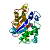
| ||||||||
|---|---|---|---|---|---|---|---|---|---|
| 1 |
| ||||||||
| Unit cell |
| ||||||||
| Details | The bilogical unit is a monomer in the asymmetric unit. |
-Components
| #1: Protein | Mass: 21227.010 Da / Num. of mol.: 1 / Source method: isolated from a natural source / Source: (natural) Momordica charantia (bitter melon) / Tissue: seed / References: UniProt: P23540, EC: 3.1.27.1 |
|---|---|
| #2: Chemical | ChemComp-U3P /   Mass: 324.181 Da / Num. of mol.: 1 / Source method: obtained synthetically / Formula: C9H13N2O9P Mass: 324.181 Da / Num. of mol.: 1 / Source method: obtained synthetically / Formula: C9H13N2O9P |
| #3: Water | ChemComp-HOH / Water Mass: 18.015 Da / Num. of mol.: 94 / Source method: isolated from a natural source / Formula: H2O Mass: 18.015 Da / Num. of mol.: 94 / Source method: isolated from a natural source / Formula: H2O |
-Experimental details
-Experiment
| Experiment | Method: X-RAY DIFFRACTION / Number of used crystals: 1 |
|---|
- Sample preparation
| Crystal | Density Matthews: 2.33 Å3/Da / Density % sol: 47.28 % | ||||||||||||||||||||||||||||||||||||
|---|---|---|---|---|---|---|---|---|---|---|---|---|---|---|---|---|---|---|---|---|---|---|---|---|---|---|---|---|---|---|---|---|---|---|---|---|---|
| Crystal grow | Temperature: 293 K / Method: vapor diffusion, hanging drop / pH: 6.7 Details: PEG 8000, sodium acetate, sodium cacodylate, pH 6.7, VAPOR DIFFUSION, HANGING DROP, temperature 293K | ||||||||||||||||||||||||||||||||||||
| Crystal grow | *PLUS | ||||||||||||||||||||||||||||||||||||
| Components of the solutions | *PLUS
|
-Data collection
| Diffraction | Mean temperature: 298 K |
|---|---|
| Diffraction source | Source: SYNCHROTRON / Site: SPring-8  / Beamline: BL44B2 / Wavelength: 0.7 Å / Beamline: BL44B2 / Wavelength: 0.7 Å |
| Detector | Type: MARRESEARCH / Detector: CCD / Date: Oct 12, 1999 / Details: mirrors |
| Radiation | Monochromator: Mirror / Protocol: SINGLE WAVELENGTH / Monochromatic (M) / Laue (L): M / Scattering type: x-ray |
| Radiation wavelength | Wavelength: 0.7 Å / Relative weight: 1 |
| Reflection | Resolution: 1.77→19.49 Å / Num. all: 28892 / Num. obs: 18370 / % possible obs: 92.7 % / Observed criterion σ(I): 3 / Redundancy: 4.3 % / Biso Wilson estimate: 15.729 Å2 / Rmerge(I) obs: 0.086 / Rsym value: 0.076 / Net I/σ(I): 6.6 |
| Reflection shell | Resolution: 1.77→1.87 Å / Redundancy: 4.2 % / Rmerge(I) obs: 0.369 / Mean I/σ(I) obs: 2 / Num. unique all: 2684 / Rsym value: 0.33 / % possible all: 94.3 |
| Reflection | *PLUS Lowest resolution: 19.4 Å / Num. measured all: 427770 |
| Reflection shell | *PLUS % possible obs: 94.3 % / Rmerge(I) obs: 0.37 |
- Processing
| Software |
| |||||||||||||||||||||||||
|---|---|---|---|---|---|---|---|---|---|---|---|---|---|---|---|---|---|---|---|---|---|---|---|---|---|---|
| Refinement | Method to determine structure: MOLECULAR REPLACEMENT Starting model: PDB ENTRY 1BK7 Resolution: 1.77→10 Å / Isotropic thermal model: isotropic / Cross valid method: THROUGHOUT / σ(F): 0 / Stereochemistry target values: Engh & Huber
| |||||||||||||||||||||||||
| Displacement parameters | Biso mean: 18.7 Å2
| |||||||||||||||||||||||||
| Refine analyze |
| |||||||||||||||||||||||||
| Refinement step | Cycle: LAST / Resolution: 1.77→10 Å
| |||||||||||||||||||||||||
| Refine LS restraints |
| |||||||||||||||||||||||||
| LS refinement shell | Resolution: 1.77→1.83 Å
| |||||||||||||||||||||||||
| Refinement | *PLUS Num. reflection obs: 18243 / % reflection Rfree: 5 % | |||||||||||||||||||||||||
| Solvent computation | *PLUS | |||||||||||||||||||||||||
| Displacement parameters | *PLUS | |||||||||||||||||||||||||
| Refine LS restraints | *PLUS
|
