 Movie
Movie Controller

[English] 日本語
 Yorodumi
- PDB-1l1r: Crystal Structure of APRTase from Giardia lamblia Complexed with ... -
+ Open data

- Basic information
| Entry | Database: PDB / ID: 1l1r | ||||||
|---|---|---|---|---|---|---|---|
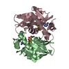
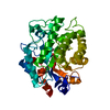
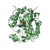
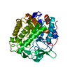
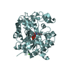
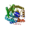
| Title | Crystal Structure of APRTase from Giardia lamblia Complexed with 9-deazaadenine, Mg2+ and PRPP | ||||||
 Components Components | Adenine phosphoribosyltransferase | ||||||
 Keywords Keywords | TRANSFERASE / APRTase / adenine / Giardia lamblia / purine metabolism / catalytic loop | ||||||
| Function / homology |  Function and homology information Function and homology informationadenine binding / adenine salvage / adenine phosphoribosyltransferase / adenine phosphoribosyltransferase activity / AMP salvage / purine ribonucleoside salvage / AMP binding / cytoplasm Similarity search - Function | ||||||
| Biological species |  Giardia intestinalis (eukaryote) Giardia intestinalis (eukaryote) | ||||||
| Method |  X-RAY DIFFRACTION / SYNCHROTRON / FOURIER SYNTHESIS / Resolution: 1.95 Å X-RAY DIFFRACTION / SYNCHROTRON / FOURIER SYNTHESIS / Resolution: 1.95 Å | ||||||
 Authors Authors | Shi, W. / Sarver, A.E. / Wang, C.C. / Tanaka, K.S. / Almo, S.C. / Schramm, V.L. | ||||||
 Citation Citation | Journal: J.Biol.Chem. / Year: 2002 Title: Closed Site Complexes of Adenine Phosphoribosyltransferase from Giardia lamblia Reveal a Mechanism of Ribosyl Migration. Authors: Shi, W. / Sarver, A.E. / Wang, C.C. / Tanaka, K.S. / Almo, S.C. / Schramm, V.L. | ||||||
| History |
|
- Structure visualization
| Structure viewer | Molecule: MolmilJmol/JSmol |
|---|
- Downloads & links
-Download
| PDBx/mmCIF format | 1l1r.cif.gz | 50.5 KB | Display | PDBx/mmCIF format |
|---|---|---|---|---|
| PDB format | pdb1l1r.ent.gz | 34.7 KB | Display | PDB format |
| PDBx/mmJSON format | 1l1r.json.gz | Tree view | PDBx/mmJSON format | |
| Others |  Other downloads Other downloads |
-Validation report
| Arichive directory | https://data.pdbj.org/pub/pdb/validation_reports/l1/1l1rftp://data.pdbj.org/pub/pdb/validation_reports/l1/1l1r | HTTPS FTP |
|---|
-Related structure data
| Related structure data | 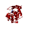 1l1qSC S: Starting model for refinement C: citing same article ( |
|---|---|
| Similar structure data |


-Links
 PDBj
PDBj
- Assembly
| Deposited unit | 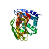
| |||||||||
|---|---|---|---|---|---|---|---|---|---|---|
| 1 | 
| |||||||||
| Unit cell |
| |||||||||
| Components on special symmetry positions |
| |||||||||
| Details | The biological assembly is a dimer generated from the monomer in the asymmetric unit by applying the crystallographic 2-fold. |
-Components
| #1: Protein | Mass: 20290.617 Da / Num. of mol.: 1 Source method: isolated from a genetically manipulated source Source: (gene. exp.) Giardia intestinalis (eukaryote) / Production host:  References: UniProt: Q967M2, adenine phosphoribosyltransferase |
|---|---|
| #2: Chemical | ChemComp-MG /   Mass: 24.305 Da / Num. of mol.: 1 / Source method: obtained synthetically / Formula: Mg Mass: 24.305 Da / Num. of mol.: 1 / Source method: obtained synthetically / Formula: Mg |
| #3: Chemical | ChemComp-9DA /   Mass: 134.139 Da / Num. of mol.: 1 / Source method: obtained synthetically / Formula: C6H6N4 Mass: 134.139 Da / Num. of mol.: 1 / Source method: obtained synthetically / Formula: C6H6N4 |
| #4: Sugar | ChemComp-PRP /   Type: D-saccharide / Mass: 390.070 Da / Num. of mol.: 1 / Source method: obtained synthetically / Formula: C5H13O14P3 Type: D-saccharide / Mass: 390.070 Da / Num. of mol.: 1 / Source method: obtained synthetically / Formula: C5H13O14P3 |
| #5: Water | ChemComp-HOH /  Mass: 18.015 Da / Num. of mol.: 79 / Source method: isolated from a natural source / Formula: H2O Mass: 18.015 Da / Num. of mol.: 79 / Source method: isolated from a natural source / Formula: H2O |
-Experimental details
-Experiment
| Experiment | Method: X-RAY DIFFRACTION / Number of used crystals: 1 |
|---|
- Sample preparation
| Crystal | Density Matthews: 2.2 Å3/Da / Density % sol: 44 % | ||||||||||||||||||||||||||||||||||||
|---|---|---|---|---|---|---|---|---|---|---|---|---|---|---|---|---|---|---|---|---|---|---|---|---|---|---|---|---|---|---|---|---|---|---|---|---|---|
| Crystal grow | Temperature: 291 K / Method: vapor diffusion, hanging drop / pH: 4.6 Details: PEG 4000, ammonium acetate, urea, sodium acetate, pH 4.6, VAPOR DIFFUSION, HANGING DROP, temperature 291K | ||||||||||||||||||||||||||||||||||||
| Crystal grow | *PLUS Temperature: 18 ℃ / Method: vapor diffusion | ||||||||||||||||||||||||||||||||||||
| Components of the solutions | *PLUS
|
-Data collection
| Diffraction | Mean temperature: 100 K |
|---|---|
| Diffraction source | Source: SYNCHROTRON / Site: NSLS  / Beamline: X9B / Wavelength: 0.98 Å / Beamline: X9B / Wavelength: 0.98 Å |
| Detector | Type: ADSC QUANTUM 4 / Detector: CCD / Date: Jun 7, 2001 |
| Radiation | Protocol: SINGLE WAVELENGTH / Monochromatic (M) / Laue (L): M / Scattering type: x-ray |
| Radiation wavelength | Wavelength: 0.98 Å / Relative weight: 1 |
| Reflection | Resolution: 1.95→25 Å / Num. all: 14082 / Num. obs: 14082 / % possible obs: 99.5 % / Observed criterion σ(F): 0 / Observed criterion σ(I): 0 / Redundancy: 5 % / Biso Wilson estimate: 12.8 Å2 / Rsym value: 0.037 / Net I/σ(I): 23.4 |
| Reflection shell | Resolution: 1.95→2.02 Å / Redundancy: 4.8 % / Mean I/σ(I) obs: 3 / Num. unique all: 1387 / Rsym value: 0.369 / % possible all: 99.8 |
| Reflection | *PLUS Num. measured all: 70505 / Rmerge(I) obs: 0.037 |
| Reflection shell | *PLUS % possible obs: 99.8 % / Rmerge(I) obs: 0.369 |
- Processing
| Software |
| |||||||||||||||||||||||||
|---|---|---|---|---|---|---|---|---|---|---|---|---|---|---|---|---|---|---|---|---|---|---|---|---|---|---|
| Refinement | Method to determine structure: FOURIER SYNTHESIS Starting model: PDB ENTRY 1L1Q Resolution: 1.95→25 Å / Rfactor Rfree error: 0.007 / Isotropic thermal model: RESTRAINED / Cross valid method: THROUGHOUT / σ(F): 2 / Stereochemistry target values: Engh & Huber
| |||||||||||||||||||||||||
| Solvent computation | Solvent model: flat model / Bsol: 37.9291 Å2 / ksol: 0.340042 e/Å3 | |||||||||||||||||||||||||
| Displacement parameters | Biso mean: 30.5 Å2
| |||||||||||||||||||||||||
| Refine analyze |
| |||||||||||||||||||||||||
| Refinement step | Cycle: LAST / Resolution: 1.95→25 Å
| |||||||||||||||||||||||||
| Refine LS restraints |
| |||||||||||||||||||||||||
| LS refinement shell | Resolution: 1.95→2.07 Å / Rfactor Rfree error: 0.022 / Total num. of bins used: 6
| |||||||||||||||||||||||||
| Refinement | *PLUS Lowest resolution: 25 Å | |||||||||||||||||||||||||
| Solvent computation | *PLUS | |||||||||||||||||||||||||
| Displacement parameters | *PLUS | |||||||||||||||||||||||||
| Refine LS restraints | *PLUS
|
