 Movie
Movie Controller

[English] 日本語
 Yorodumi
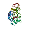
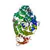
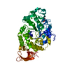
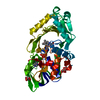
- PDB-1ua7: Crystal Structure Analysis of Alpha-Amylase from Bacillus Subtili... -
+ Open data

- Basic information
| Entry | Database: PDB / ID: 1ua7 | ||||||||||||
|---|---|---|---|---|---|---|---|---|---|---|---|---|---|
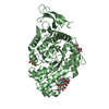
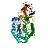
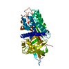
| Title | Crystal Structure Analysis of Alpha-Amylase from Bacillus Subtilis complexed with Acarbose | ||||||||||||
 Components Components | Alpha-amylase | ||||||||||||
 Keywords Keywords | HYDROLASE / BETA-ALPHA-BARRELS / ACARBOSE / GREEK-KEY MOTIF | ||||||||||||
| Function / homology |  Function and homology information Function and homology informationalpha-amylase / alpha-amylase activity / carbohydrate metabolic process / extracellular region / metal ion binding Similarity search - Function | ||||||||||||
| Biological species |  | ||||||||||||
| Method |  X-RAY DIFFRACTION / FOURIER SYNTHESIS / Resolution: 2.21 Å X-RAY DIFFRACTION / FOURIER SYNTHESIS / Resolution: 2.21 Å | ||||||||||||
 Authors Authors | Kagawa, M. / Fujimoto, Z. / Momma, M. / Takase, K. / Mizuno, H. | ||||||||||||
 Citation Citation | Journal: J.BACTERIOL. / Year: 2003 Title: Crystal structure of Bacillus subtilis alpha-amylase in complex with acarbose Authors: Kagawa, M. / Fujimoto, Z. / Momma, M. / Takase, K. / Mizuno, H. #1: Journal: J.Mol.Biol. / Year: 1998Title: Crystal structure of a catalytic-site mutant alpha-amylase from Bacillus subtilis complexed with maltopentaose Authors: Fujimoto, Z. / Takase, K. / Doui, N. / Momma, M. / Matsumoto, T. / Mizuno, H. #2: Journal: J.Mol.Biol. / Year: 1993Title: Crystallization and preliminary X-ray studies of wild type and catalytic-site mutant alpha-amylase from Bacillus subtilis Authors: Mizuno, H. / Morimoto, Y. / Tsukihara, T. / Matsumoto, T. / Takase, K. #3: Journal: BIOCHIM.BIOPHYS.ACTA / Year: 1992Title: Site-directed mutagenesis of active site residues in Bacillus subtilis alpha-amylase Authors: Takase, K. / Matsumoto, T. / Mizuno, H. / Yamane, K. #4: Journal: J.BIOCHEM.(TOKYO) / Year: 1984Title: Changes in the properties and molecular weights of Bacillus subtilis M-type and N-type alpha-amylases resulting from a spontaneous deletion Authors: Yamane, K. / Hirata, Y. / Furusato, T. / Yamazaki, H. / Nakayama, A. | ||||||||||||
| History |
|
- Structure visualization
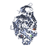
| Structure viewer | Molecule: MolmilJmol/JSmol |
|---|
- Downloads & links
-Download
| PDBx/mmCIF format | 1ua7.cif.gz | 111.1 KB | Display | PDBx/mmCIF format |
|---|---|---|---|---|
| PDB format | pdb1ua7.ent.gz | 81.5 KB | Display | PDB format |
| PDBx/mmJSON format | 1ua7.json.gz | Tree view | PDBx/mmJSON format | |
| Others |  Other downloads Other downloads |
-Validation report
| Summary document | 1ua7_validation.pdf.gz | 1 MB | Display | wwPDB validaton report |
|---|---|---|---|---|
| Full document | 1ua7_full_validation.pdf.gz | 1 MB | Display | |
| Data in XML | 1ua7_validation.xml.gz | 22.6 KB | Display | |
| Data in CIF | 1ua7_validation.cif.gz | 34.2 KB | Display | |
| Arichive directory | https://data.pdbj.org/pub/pdb/validation_reports/ua/1ua7ftp://data.pdbj.org/pub/pdb/validation_reports/ua/1ua7 | HTTPS FTP |
-Related structure data
| Related structure data | |
|---|---|
| Similar structure data |


-Links
 PDBj
PDBj

- Assembly
| Deposited unit | 
| ||||||||
|---|---|---|---|---|---|---|---|---|---|
| 1 |
| ||||||||
| Unit cell |
|
-Components
-Protein , 1 types, 1 molecules A
| #1: Protein | Mass: 46796.984 Da / Num. of mol.: 1 / Fragment: residues 4-425 / Mutation: N356Q Source method: isolated from a genetically manipulated source Source: (gene. exp.) |
|---|
-Sugars , 2 types, 2 molecules
| #2: Polysaccharide | 4,6-dideoxy-alpha-D-xylo-hexopyranose-(1-4)-alpha-D-glucopyranose Source method: isolated from a genetically manipulated source |
|---|---|
| #3: Polysaccharide | alpha-D-quinovopyranose-(1-4)-beta-D-glucopyranose Source method: isolated from a genetically manipulated source |
-Non-polymers , 3 types, 442 molecules 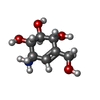


| #4: Chemical |  Mass: 175.182 Da / Num. of mol.: 2 Mass: 175.182 Da / Num. of mol.: 2Source method: isolated from a genetically manipulated source Formula: C7H13NO4 / Comment: antibiotic*YM #5: Chemical |  Mass: 40.078 Da / Num. of mol.: 3 / Source method: obtained synthetically / Formula: Ca Mass: 40.078 Da / Num. of mol.: 3 / Source method: obtained synthetically / Formula: Ca#6: Water | ChemComp-HOH / | Mass: 18.015 Da / Num. of mol.: 437 / Source method: isolated from a natural source / Formula: H2O |
|---|
-Experimental details
-Experiment
| Experiment | Method: X-RAY DIFFRACTION / Number of used crystals: 1 |
|---|
- Sample preparation
| Crystal | Density Matthews: 2.83 Å3/Da / Density % sol: 56.22 % |
|---|---|
| Crystal grow | Temperature: 293 K / Method: vapor diffusion, hanging drop / pH: 7.2 Details: PEG 3350, calcium chloride, Tris-HCl, acarbose, pH 7.2, VAPOR DIFFUSION, HANGING DROP, temperature 293.0K |
-Data collection
| Diffraction | Mean temperature: 100 K |
|---|---|
| Diffraction source | Source: ROTATING ANODE / Type: RIGAKU ULTRAX 18 / Wavelength: 1.5418 Å |
| Detector | Type: RIGAKU RAXIS IV / Detector: IMAGE PLATE / Date: Feb 1, 2001 |
| Radiation | Protocol: SINGLE WAVELENGTH / Monochromatic (M) / Laue (L): M / Scattering type: x-ray |
| Radiation wavelength | Wavelength: 1.5418 Å / Relative weight: 1 |
| Reflection | Resolution: 2.2→45.49 Å / Num. all: 31194 / Num. obs: 31194 / % possible obs: 93.7 % / Redundancy: 3.46 % / Biso Wilson estimate: 6.5 Å2 / Rmerge(I) obs: 0.097 / Net I/σ(I): 22.9 |
| Reflection shell | Resolution: 2.2→2.28 Å / Redundancy: 3.4 % / Rmerge(I) obs: 0.239 / Mean I/σ(I) obs: 7.7 / Num. unique all: 3057 / % possible all: 93.7 |
- Processing
| Software | Name: CNS / Version: 1 / Classification: refinement | |||||||||||||||||||||||||
|---|---|---|---|---|---|---|---|---|---|---|---|---|---|---|---|---|---|---|---|---|---|---|---|---|---|---|
| Refinement | Method to determine structure: FOURIER SYNTHESIS / Resolution: 2.21→19.97 Å / Rfactor Rfree error: 0.005 / Isotropic thermal model: RESTRAINED / Cross valid method: THROUGHOUT / σ(F): 0
| |||||||||||||||||||||||||
| Solvent computation | Solvent model: FLAT MODEL / Bsol: 56.5617 Å2 / ksol: 0.346066 e/Å3 | |||||||||||||||||||||||||
| Displacement parameters | Biso mean: 21.4 Å2
| |||||||||||||||||||||||||
| Refine analyze |
| |||||||||||||||||||||||||
| Refinement step | Cycle: LAST / Resolution: 2.21→19.97 Å
| |||||||||||||||||||||||||
| Refine LS restraints |
| |||||||||||||||||||||||||
| LS refinement shell | Resolution: 2.2→2.34 Å / Rfactor Rfree error: 0.016 / Total num. of bins used: 6
| |||||||||||||||||||||||||
| Xplor file |
|
