 Movie
Movie Controller

[English] 日本語
 Yorodumi
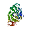
- PDB-1bag: ALPHA-AMYLASE FROM BACILLUS SUBTILIS COMPLEXED WITH MALTOPENTAOSE -
+ Open data

- Basic information
| Entry | Database: PDB / ID: 1bag | |||||||||
|---|---|---|---|---|---|---|---|---|---|---|
| Title | ALPHA-AMYLASE FROM BACILLUS SUBTILIS COMPLEXED WITH MALTOPENTAOSE | |||||||||
 Components Components | ALPHA-1,4-GLUCAN-4-GLUCANOHYDROLASE | |||||||||
 Keywords Keywords | ALPHA-AMYLASE / BACILLUS SUBTILIS / MALTOPENTAOSE / CATALYTIC-SITE MUTANT | |||||||||
| Function / homology |  Function and homology information Function and homology informationalpha-amylase / alpha-amylase activity / carbohydrate metabolic process / extracellular region / metal ion binding Similarity search - Function | |||||||||
| Biological species |  | |||||||||
| Method |  X-RAY DIFFRACTION / SYNCHROTRON / MIR / Resolution: 2.5 Å X-RAY DIFFRACTION / SYNCHROTRON / MIR / Resolution: 2.5 Å | |||||||||
 Authors Authors | Fujimoto, Z. / Mizuno, H. / Takase, K. / Doui, N. | |||||||||
 Citation Citation | Journal: J.Mol.Biol. / Year: 1998 Title: Crystal structure of a catalytic-site mutant alpha-amylase from Bacillus subtilis complexed with maltopentaose. Authors: Fujimoto, Z. / Takase, K. / Doui, N. / Momma, M. / Matsumoto, T. / Mizuno, H. #1: Journal: J.Mol.Biol. / Year: 1993Title: Crystallization and Preliminary X-Ray Studies of Wild Type and Catalytic-Site Mutant Alpha-Amylase from Bacillus Subtilis Authors: Mizuno, H. / Morimoto, Y. / Tsukihara, T. / Matsumoto, T. / Takase, K. #2: Journal: Biochim.Biophys.Acta / Year: 1992Title: Site-Directed Mutagenesis of Active Site Residues in Bacillus Subtilis Alpha-Amylase Authors: Takase, K. / Matsumoto, T. / Mizuno, H. / Yamane, K. #3: Journal: J.Biochem.(Tokyo) / Year: 1984Title: Changes in the Properties and Molecular Weights of Bacillus Subtilis M-Type and N-Type Alpha-Amylases Resulting from a Spontaneous Deletion Authors: Yamane, K. / Hirata, Y. / Furusato, T. / Yamazaki, H. / Nakayama, A. | |||||||||
| History |
|
- Structure visualization
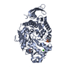
| Structure viewer | Molecule: MolmilJmol/JSmol |
|---|
- Downloads & links
-Download
| PDBx/mmCIF format | 1bag.cif.gz | 100.9 KB | Display | PDBx/mmCIF format |
|---|---|---|---|---|
| PDB format | pdb1bag.ent.gz | 75.6 KB | Display | PDB format |
| PDBx/mmJSON format | 1bag.json.gz | Tree view | PDBx/mmJSON format | |
| Others |  Other downloads Other downloads |
-Validation report
| Arichive directory | https://data.pdbj.org/pub/pdb/validation_reports/ba/1bagftp://data.pdbj.org/pub/pdb/validation_reports/ba/1bag | HTTPS FTP |
|---|
-Related structure data
| Similar structure data |
|---|


-Links
 PDBj
PDBj

- Assembly
| Deposited unit | 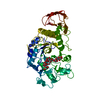
| ||||||||
|---|---|---|---|---|---|---|---|---|---|
| 1 |
| ||||||||
| Unit cell |
|
-Components
| #1: Protein | Mass: 47067.309 Da / Num. of mol.: 1 / Mutation: E208Q Source method: isolated from a genetically manipulated source Details: COMPLEXED WITH MALTOPENTAOSE / Source: (gene. exp.) | ||
|---|---|---|---|
| #2: Polysaccharide | alpha-D-glucopyranose-(1-4)-alpha-D-glucopyranose-(1-4)-alpha-D-glucopyranose-(1-4)-alpha-D- ...alpha-D-glucopyranose-(1-4)-alpha-D-glucopyranose-(1-4)-alpha-D-glucopyranose-(1-4)-alpha-D-glucopyranose-(1-4)-beta-D-glucopyranose Source method: isolated from a genetically manipulated source | ||
| #3: Chemical |   Mass: 40.078 Da / Num. of mol.: 3 / Source method: obtained synthetically / Formula: Ca Mass: 40.078 Da / Num. of mol.: 3 / Source method: obtained synthetically / Formula: Ca#4: Water | ChemComp-HOH / |  Mass: 18.015 Da / Num. of mol.: 146 / Source method: isolated from a natural source / Formula: H2O Mass: 18.015 Da / Num. of mol.: 146 / Source method: isolated from a natural source / Formula: H2O |
-Experimental details
-Experiment
| Experiment | Method: X-RAY DIFFRACTION / Number of used crystals: 4 |
|---|
- Sample preparation
| Crystal | Density Matthews: 3.2 Å3/Da / Density % sol: 62 % | ||||||||||||||||||||||||||||||||||||||||||
|---|---|---|---|---|---|---|---|---|---|---|---|---|---|---|---|---|---|---|---|---|---|---|---|---|---|---|---|---|---|---|---|---|---|---|---|---|---|---|---|---|---|---|---|
| Crystal grow | pH: 7.5 Details: PROTEIN WAS CRYSTALLIZED FROM 10% PEG 3350, 3.5 MM CALCIUM CHLORIDE, 10 MM TRIS/HCL, PH 7.5. | ||||||||||||||||||||||||||||||||||||||||||
| Crystal | *PLUS | ||||||||||||||||||||||||||||||||||||||||||
| Crystal grow | *PLUS Method: vapor diffusion, hanging drop | ||||||||||||||||||||||||||||||||||||||||||
| Components of the solutions | *PLUS
|
-Data collection
| Diffraction | Mean temperature: 288 K |
|---|---|
| Diffraction source | Source: SYNCHROTRON / Site: Photon Factory  / Beamline: BL-6A / Wavelength: 1 / Beamline: BL-6A / Wavelength: 1 |
| Detector | Type: RIGAKU / Detector: IMAGE PLATE / Date: Oct 1, 1995 / Details: COLLIMATOR |
| Radiation | Monochromatic (M) / Laue (L): M / Scattering type: x-ray |
| Radiation wavelength | Wavelength: 1 Å / Relative weight: 1 |
| Reflection | Resolution: 2.5→100 Å / Num. obs: 19373 / % possible obs: 80.4 % / Observed criterion σ(I): 2 / Redundancy: 2.5 % / Biso Wilson estimate: 12.2 Å2 / Rmerge(I) obs: 0.07 / Net I/σ(I): 16 |
| Reflection shell | Resolution: 2.5→2.61 Å / Redundancy: 2.5 % / Rmerge(I) obs: 0.19 / Mean I/σ(I) obs: 5 / % possible all: 44 |
| Reflection | *PLUS Num. obs: 18097 / Num. measured all: 56354 / Rmerge(I) obs: 0.067 |
- Processing
| Software |
| ||||||||||||||||||||||||||||||||||||||||||||||||||||||||||||||||||||||||||||||||
|---|---|---|---|---|---|---|---|---|---|---|---|---|---|---|---|---|---|---|---|---|---|---|---|---|---|---|---|---|---|---|---|---|---|---|---|---|---|---|---|---|---|---|---|---|---|---|---|---|---|---|---|---|---|---|---|---|---|---|---|---|---|---|---|---|---|---|---|---|---|---|---|---|---|---|---|---|---|---|---|---|---|
| Refinement | Method to determine structure: MIR / Resolution: 2.5→7 Å / Rfactor Rfree error: 0.006 / Data cutoff high absF: 10000000 / Data cutoff low absF: 0.001 / Isotropic thermal model: RESTRAINED / Cross valid method: THROUGHOUT / σ(F): 2
| ||||||||||||||||||||||||||||||||||||||||||||||||||||||||||||||||||||||||||||||||
| Displacement parameters | Biso mean: 15.9 Å2 | ||||||||||||||||||||||||||||||||||||||||||||||||||||||||||||||||||||||||||||||||
| Refine analyze |
| ||||||||||||||||||||||||||||||||||||||||||||||||||||||||||||||||||||||||||||||||
| Refinement step | Cycle: LAST / Resolution: 2.5→7 Å
| ||||||||||||||||||||||||||||||||||||||||||||||||||||||||||||||||||||||||||||||||
| Refine LS restraints |
| ||||||||||||||||||||||||||||||||||||||||||||||||||||||||||||||||||||||||||||||||
| LS refinement shell | Resolution: 2.5→2.65 Å / Rfactor Rfree error: 0.027 / Total num. of bins used: 6
| ||||||||||||||||||||||||||||||||||||||||||||||||||||||||||||||||||||||||||||||||
| Xplor file |
| ||||||||||||||||||||||||||||||||||||||||||||||||||||||||||||||||||||||||||||||||
| Software | *PLUS Name: X-PLOR / Version: 3.8 / Classification: refinement | ||||||||||||||||||||||||||||||||||||||||||||||||||||||||||||||||||||||||||||||||
| Refine LS restraints | *PLUS
|
