 Movie
Movie Controller

[English] 日本語
 Yorodumi
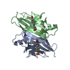
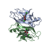
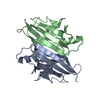
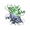
- PDB-1tha: MECHANISM OF MOLECULAR RECOGNITION. STRUCTURAL ASPECTS OF 3,3'-DI... -
+ Open data

- Basic information
| Entry | Database: PDB / ID: 1tha | ||||||
|---|---|---|---|---|---|---|---|
| Title | MECHANISM OF MOLECULAR RECOGNITION. STRUCTURAL ASPECTS OF 3,3'-DIIODO-L-THYRONINE BINDING TO HUMAN SERUM TRANSTHYRETIN | ||||||
 Components Components | TRANSTHYRETIN | ||||||
 Keywords Keywords | TRANSPORT(THYROXINE) | ||||||
| Function / homology |  Function and homology information Function and homology informationRetinoid cycle disease events / thyroid hormone binding / The canonical retinoid cycle in rods (twilight vision) / Non-integrin membrane-ECM interactions / purine nucleobase metabolic process / Retinoid metabolism and transport / hormone activity / azurophil granule lumen / Amyloid fiber formation / Neutrophil degranulation ...Retinoid cycle disease events / thyroid hormone binding / The canonical retinoid cycle in rods (twilight vision) / Non-integrin membrane-ECM interactions / purine nucleobase metabolic process / Retinoid metabolism and transport / hormone activity / azurophil granule lumen / Amyloid fiber formation / Neutrophil degranulation / extracellular space / extracellular exosome / extracellular region / identical protein bindingSimilarity search - Function | ||||||
| Biological species |  Homo sapiens (human) Homo sapiens (human) | ||||||
| Method | X-RAY DIFFRACTION / Resolution: 2 Å | ||||||
 Authors Authors | Wojtczak, A. / Luft, J. / Cody, V. | ||||||
 Citation Citation | Journal: J.Biol.Chem. / Year: 1992 Title: Mechanism of molecular recognition. Structural aspects of 3,3'-diiodo-L-thyronine binding to human serum transthyretin. Authors: Wojtczak, A. / Luft, J. / Cody, V. #1: Journal: Nature / Year: 1977Title: Protein-DNA and Protein-Hormone Interactions in Prealbumin, a Model of the Thyroid Hormone Nuclear Receptor (Query) Authors: Blake, C.C.F. / Oatley, S.J. #4: Journal: J.Mol.Biol. / Year: 1974Title: Structure of Human Plasma Prealbumin at 2.5 Angstroms Resolution, a Preliminary Report on the Polypeptide Chain Conformation, Quaternary Structure and Thyroxine Binding Authors: Blake, C.C.F. / Geisow, M.J. / Swan, I.D.A. / Rerat, C. / Rerat, B. #5: Journal: J.Mol.Biol. / Year: 1971Title: An X-Ray Study of the Subunit Structure of Prealbumin Authors: Blake, C.C.F. / Swan, I.D.A. / Rerat, C. / Berthou, J. / Laurent, A. / Rerat, B. | ||||||
| History |
| ||||||
| Remark 700 | SHEET THE FIRST STRAND OF THE *EXTERNAL* SHEET IN EACH SUBUNIT IS DISCONTINUOUS. TO ACCOMMODATE ...SHEET THE FIRST STRAND OF THE *EXTERNAL* SHEET IN EACH SUBUNIT IS DISCONTINUOUS. TO ACCOMMODATE THESE DISCONTINUITIES EACH *EXTERNAL* SHEET IS REPRESENTED HERE TWICE, WITH A DIFFERENT STRAND 1 IN EACH CASE. STRANDS 2, 3, 4 OF *X1A* ARE IDENTICAL TO STRANDS 2, 3, 4 OF *X2A*. SIMILARLY STRANDS 2, 3, 4 OF *X1B* ARE IDENTICAL TO STRANDS 2, 3, 4 OF *X2B*. THIS DESCRIPTION IS CONSISTENT WITH THAT USED BY BLAKE AND COWORKERS FOR THE NATIVE TRANSTHYRETIN IN PROTEIN DATA BANK ENTRY 2PAB. |
- Structure visualization
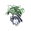
| Structure viewer | Molecule: MolmilJmol/JSmol |
|---|
- Downloads & links
-Download
| PDBx/mmCIF format | 1tha.cif.gz | 61.7 KB | Display | PDBx/mmCIF format |
|---|---|---|---|---|
| PDB format | pdb1tha.ent.gz | 45.5 KB | Display | PDB format |
| PDBx/mmJSON format | 1tha.json.gz | Tree view | PDBx/mmJSON format | |
| Others |  Other downloads Other downloads |
-Validation report
| Arichive directory | https://data.pdbj.org/pub/pdb/validation_reports/th/1thaftp://data.pdbj.org/pub/pdb/validation_reports/th/1tha | HTTPS FTP |
|---|
-Related structure data
| Similar structure data |
|---|


-Links
 PDBj
PDBj


- Assembly
| Deposited unit | 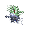
| |||||||||
|---|---|---|---|---|---|---|---|---|---|---|
| 1 | 
| |||||||||
| Unit cell |
| |||||||||
| Components on special symmetry positions |
|
-Components
| #1: Protein | Mass: 13776.376 Da / Num. of mol.: 2 Source method: isolated from a genetically manipulated source Source: (gene. exp.) Homo sapiens (human) / References: UniProt: P02766#2: Chemical | 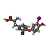  Mass: 525.077 Da / Num. of mol.: 2 / Source method: obtained synthetically / Formula: C15H13I2NO4 Mass: 525.077 Da / Num. of mol.: 2 / Source method: obtained synthetically / Formula: C15H13I2NO4#3: Water | ChemComp-HOH / | Water Mass: 18.015 Da / Num. of mol.: 98 / Source method: isolated from a natural source / Formula: H2O Mass: 18.015 Da / Num. of mol.: 98 / Source method: isolated from a natural source / Formula: H2OSequence details | SEQUENCE ADVISORY NOTICE: DIFFERENCE BETWEEN SWISS-PROT AND PDB SEQUENCE. SWISS-PROT ENTRY NAME: ...SEQUENCE ADVISORY NOTICE: DIFFERENCE | |
|---|
-Experimental details
-Experiment
| Experiment | Method: X-RAY DIFFRACTION |
|---|
- Sample preparation
| Crystal | Density Matthews: 2.27 Å3/Da / Density % sol: 45.72 % |
|---|---|
| Crystal grow | *PLUS Method: vapor diffusion, hanging drop |
| Components of the solutions | *PLUS Common name: ammonium sulfate |
-Data collection
| Reflection | *PLUS Highest resolution: 1.93 Å / Num. obs: 12244 / Observed criterion σ(I): 2 / Num. measured all: 38040 / Rmerge(I) obs: 0.073 |
|---|
- Processing
| Software |
| ||||||||||||||||||||||||||||||||||||||||||||||||||||||||||||||||||||||||||||||||
|---|---|---|---|---|---|---|---|---|---|---|---|---|---|---|---|---|---|---|---|---|---|---|---|---|---|---|---|---|---|---|---|---|---|---|---|---|---|---|---|---|---|---|---|---|---|---|---|---|---|---|---|---|---|---|---|---|---|---|---|---|---|---|---|---|---|---|---|---|---|---|---|---|---|---|---|---|---|---|---|---|---|
| Refinement | Resolution: 2→8 Å / Rfactor obs: 0.185 Details: THIS COORDINATE SET COMPRISES TWO CHAINS REPRESENTING TWO CHEMICALLY EQUIVALENT, BUT CRYSTALLOGRAPHICALLY DISTINCT, ENTITIES. THE OTHER HALF OF THE COMPLETE TETRAMER MAY BE GENERATED FROM ...Details: THIS COORDINATE SET COMPRISES TWO CHAINS REPRESENTING TWO CHEMICALLY EQUIVALENT, BUT CRYSTALLOGRAPHICALLY DISTINCT, ENTITIES. THE OTHER HALF OF THE COMPLETE TETRAMER MAY BE GENERATED FROM THIS DIMER BY THE APPLICATION OF THE CRYSTALLOGRAPHIC DIAD PARALLEL TO Z THROUGH THE ORIGIN OF THIS COORDINATE SYSTEM, I. E. XPRIME=-X, YPRIME=-Y, ZPRIME=Z. | ||||||||||||||||||||||||||||||||||||||||||||||||||||||||||||||||||||||||||||||||
| Refinement step | Cycle: LAST / Resolution: 2→8 Å
| ||||||||||||||||||||||||||||||||||||||||||||||||||||||||||||||||||||||||||||||||
| Refine LS restraints |
| ||||||||||||||||||||||||||||||||||||||||||||||||||||||||||||||||||||||||||||||||
| Refinement | *PLUS Highest resolution: 2 Å / Lowest resolution: 8 Å / Num. reflection obs: 12215 / σ(F): 3 / Rfactor obs: 0.185 | ||||||||||||||||||||||||||||||||||||||||||||||||||||||||||||||||||||||||||||||||
| Solvent computation | *PLUS | ||||||||||||||||||||||||||||||||||||||||||||||||||||||||||||||||||||||||||||||||
| Displacement parameters | *PLUS | ||||||||||||||||||||||||||||||||||||||||||||||||||||||||||||||||||||||||||||||||
| Refine LS restraints | *PLUS
|
