 Movie
Movie Controller

[English] 日本語
 Yorodumi
- PDB-1tf6: CO-CRYSTAL STRUCTURE OF XENOPUS TFIIIA ZINC FINGER DOMAIN BOUND T... -
+ Open data

- Basic information
| Entry | Database: PDB / ID: 1tf6 | ||||||
|---|---|---|---|---|---|---|---|
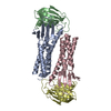
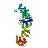
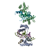
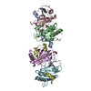
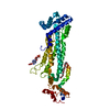
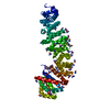
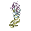
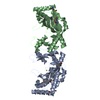
| Title | CO-CRYSTAL STRUCTURE OF XENOPUS TFIIIA ZINC FINGER DOMAIN BOUND TO THE 5S RIBOSOMAL RNA GENE INTERNAL CONTROL REGION | ||||||
 Components Components |
| ||||||
 Keywords Keywords | TRANSCRIPTION/DNA / COMPLEX (TRANSCRIPTION REGULATION-DNA) / RNA POLYMERASE III / TRANSCRIPTION INITIATION / ZINC FINGER PROTEIN / TRANSCRIPTION-DNA COMPLEX | ||||||
| Function / homology |  Function and homology information Function and homology informationribosomal large subunit biogenesis / DNA binding / RNA binding / zinc ion binding / nucleus Similarity search - Function | ||||||
| Biological species | |||||||
| Method |  X-RAY DIFFRACTION / SYNCHROTRON / MIR / Resolution: 3.1 Å X-RAY DIFFRACTION / SYNCHROTRON / MIR / Resolution: 3.1 Å | ||||||
 Authors Authors | Nolte, R.T. / Conlin, R.M. / Harrison, S.C. / Brown, R.S. | ||||||
 Citation Citation | Journal: Proc.Natl.Acad.Sci.USA / Year: 1998 Title: Differing roles for zinc fingers in DNA recognition: structure of a six-finger transcription factor IIIA complex. Authors: Nolte, R.T. / Conlin, R.M. / Harrison, S.C. / Brown, R.S. | ||||||
| History |
|
- Structure visualization
| Structure viewer | Molecule: MolmilJmol/JSmol |
|---|
- Downloads & links
-Download
| PDBx/mmCIF format | 1tf6.cif.gz | 154.9 KB | Display | PDBx/mmCIF format |
|---|---|---|---|---|
| PDB format | pdb1tf6.ent.gz | 116.7 KB | Display | PDB format |
| PDBx/mmJSON format | 1tf6.json.gz | Tree view | PDBx/mmJSON format | |
| Others |  Other downloads Other downloads |
-Validation report
| Arichive directory | https://data.pdbj.org/pub/pdb/validation_reports/tf/1tf6ftp://data.pdbj.org/pub/pdb/validation_reports/tf/1tf6 | HTTPS FTP |
|---|
-Related structure data
| Similar structure data |
|---|

-Links
 PDBj
PDBj


- Assembly
| Deposited unit | 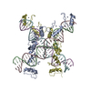
| ||||||||||||||||||||
|---|---|---|---|---|---|---|---|---|---|---|---|---|---|---|---|---|---|---|---|---|---|
| 1 | 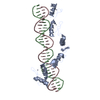
| ||||||||||||||||||||
| 2 | 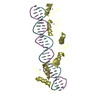
| ||||||||||||||||||||
| Unit cell |
| ||||||||||||||||||||
| Noncrystallographic symmetry (NCS) | NCS oper:
|
-Components
| #1: DNA chain | Mass: 9634.182 Da / Num. of mol.: 2 / Fragment: INTERNAL PROMOTER REGION / Source method: obtained synthetically #2: DNA chain | Mass: 9434.062 Da / Num. of mol.: 2 / Fragment: INTERNAL PROMOTER REGION / Source method: obtained synthetically #3: Protein | Mass: 22126.504 Da / Num. of mol.: 2 / Fragment: NH2-TERMINAL SIX FINGERS, RESIDUE 1-190 Source method: isolated from a genetically manipulated source Source: (gene. exp.)  #4: Chemical | ChemComp-ZN /   Mass: 65.409 Da / Num. of mol.: 12 / Source method: obtained synthetically / Formula: Zn Mass: 65.409 Da / Num. of mol.: 12 / Source method: obtained synthetically / Formula: Zn |
|---|
-Experimental details
-Experiment
| Experiment | Method: X-RAY DIFFRACTION / Number of used crystals: 3 |
|---|
- Sample preparation
| Crystal | Density Matthews: 3.8 Å3/Da / Density % sol: 70 % Description: DATA WERE FILTERED LOCALLY WITH AN I/SIGMA(I) CUTOFF OF 1.0 - ELIMINATION OF UNRELIABLE MEASUREMENTS. | ||||||||||||||||||||||||||||||||||||||||||||||||
|---|---|---|---|---|---|---|---|---|---|---|---|---|---|---|---|---|---|---|---|---|---|---|---|---|---|---|---|---|---|---|---|---|---|---|---|---|---|---|---|---|---|---|---|---|---|---|---|---|---|
| Crystal grow | Temperature: 291 K / Method: vapor diffusion, hanging drop / pH: 8 Details: COMPLEX WAS CRYSTALLIZED FROM 22.5% PEG 4000, 165 MM NACL, 35 MM SODIUM ACETATE, 3.2 MM DTT, 9.2% (VOL/VOL) GLYCEROL, 1.8 MM NAN3, 1.8 MM CADAVERINE-2HCL, 5.5 MM TRIS-HCL, PH 8.0, VAPOR ...Details: COMPLEX WAS CRYSTALLIZED FROM 22.5% PEG 4000, 165 MM NACL, 35 MM SODIUM ACETATE, 3.2 MM DTT, 9.2% (VOL/VOL) GLYCEROL, 1.8 MM NAN3, 1.8 MM CADAVERINE-2HCL, 5.5 MM TRIS-HCL, PH 8.0, VAPOR DIFFUSION, HANGING DROP, temperature 291.00K | ||||||||||||||||||||||||||||||||||||||||||||||||
| Components of the solutions |
|
-Data collection
| Diffraction | Mean temperature: 113 K |
|---|---|
| Diffraction source | Source: SYNCHROTRON / Site: NSLS  / Beamline: X12B / Beamline: X12B |
| Detector | Type: MARRESEARCH / Detector: IMAGE PLATE / Date: Oct 1, 1996 / Details: MIRRORS |
| Radiation | Monochromator: SI(111) / Protocol: SINGLE WAVELENGTH / Monochromatic (M) / Laue (L): M / Scattering type: x-ray |
| Radiation wavelength | Relative weight: 1 |
| Reflection | Resolution: 3→25 Å / Num. obs: 19034 / % possible obs: 77.4 % / Observed criterion σ(I): -3 / Redundancy: 6.7 % / Biso Wilson estimate: 39.7 Å2 / Rmerge(I) obs: 0.128 / Rsym value: 0.06 |
| Reflection | *PLUS Highest resolution: 3 Å / Lowest resolution: 25 Å / % possible obs: 77.4 % / Num. measured all: 126746 |
- Processing
| Software |
| ||||||||||||||||||||||||||||||||||||||||||||||||||||||||||||||||||||||||||||||||
|---|---|---|---|---|---|---|---|---|---|---|---|---|---|---|---|---|---|---|---|---|---|---|---|---|---|---|---|---|---|---|---|---|---|---|---|---|---|---|---|---|---|---|---|---|---|---|---|---|---|---|---|---|---|---|---|---|---|---|---|---|---|---|---|---|---|---|---|---|---|---|---|---|---|---|---|---|---|---|---|---|---|
| Refinement | Method to determine structure: MIR / Resolution: 3.1→8 Å / Rfactor Rfree error: 0.016 / Data cutoff high absF: 1000000 / Data cutoff low absF: 0 / Isotropic thermal model: RESTRAINED / Cross valid method: THROUGHOUT / σ(F): 2 Details: ISOTROPIC B FACTORS WERE REFINED AGAINST UNSHARPENED NATIVE DATA WHICH HAS AN INHERENT B FACTOR OF 65.0, AND R-FACTORS WERE CALCULATED USING ANISOTROPICALLY SHARPENED DATA.
| ||||||||||||||||||||||||||||||||||||||||||||||||||||||||||||||||||||||||||||||||
| Displacement parameters | Biso mean: 65 Å2
| ||||||||||||||||||||||||||||||||||||||||||||||||||||||||||||||||||||||||||||||||
| Refine analyze |
| ||||||||||||||||||||||||||||||||||||||||||||||||||||||||||||||||||||||||||||||||
| Refinement step | Cycle: LAST / Resolution: 3.1→8 Å
| ||||||||||||||||||||||||||||||||||||||||||||||||||||||||||||||||||||||||||||||||
| Refine LS restraints |
| ||||||||||||||||||||||||||||||||||||||||||||||||||||||||||||||||||||||||||||||||
| Refine LS restraints NCS | NCS model details: NONE | ||||||||||||||||||||||||||||||||||||||||||||||||||||||||||||||||||||||||||||||||
| Xplor file |
| ||||||||||||||||||||||||||||||||||||||||||||||||||||||||||||||||||||||||||||||||
| Software | *PLUS Name: X-PLOR / Version: 3.851 / Classification: refinement | ||||||||||||||||||||||||||||||||||||||||||||||||||||||||||||||||||||||||||||||||
| Refinement | *PLUS Highest resolution: 3.1 Å / Lowest resolution: 8 Å / σ(F): 2 / % reflection Rfree: 2.9 % | ||||||||||||||||||||||||||||||||||||||||||||||||||||||||||||||||||||||||||||||||
| Solvent computation | *PLUS | ||||||||||||||||||||||||||||||||||||||||||||||||||||||||||||||||||||||||||||||||
| Displacement parameters | *PLUS Biso mean: 65 Å2 | ||||||||||||||||||||||||||||||||||||||||||||||||||||||||||||||||||||||||||||||||
| Refine LS restraints | *PLUS
|
