symbiont-mediated disruption of host focal adhesion / NAD+-protein-arginine ADP-ribosyltransferase / NAD+-protein-arginine ADP-ribosyltransferase activity / protein secretion by the type III secretion system / nucleotidyltransferase activity / GTPase activator activity / toxin activity / extracellular region Similarity search - Function
Single alpha-helices involved in coiled-coils or other helix-helix interfaces - #2690 / Type III secretion chaperone SycE / Bacterial Rho GTPase-activating protein (GAP) domain profile. / Type III secretion system effector protein YopE-like / Virulence factor YopE, GAP domain / Virulence factor YopE, GAP domain superfamily / Yersinia virulence determinant (YopE) / : / Tir chaperone protein (CesT) family / Tir chaperone protein (CesT) family ...Single alpha-helices involved in coiled-coils or other helix-helix interfaces - #2690 / Type III secretion chaperone SycE / Bacterial Rho GTPase-activating protein (GAP) domain profile. / Type III secretion system effector protein YopE-like / Virulence factor YopE, GAP domain / Virulence factor YopE, GAP domain superfamily / Yersinia virulence determinant (YopE) / : / Tir chaperone protein (CesT) family / Tir chaperone protein (CesT) family / Yope Regulator; Chain: A, - #10 / Yope Regulator; Chain: A, / ADP ribosyltransferase / ADP-ribosyltransferase exoenzyme / Toxin-related mono-ADP-ribosyltransferase (TR mART) core domain profile. / Single alpha-helices involved in coiled-coils or other helix-helix interfaces / Helix non-globular / Special / 2-Layer Sandwich / Alpha Beta Similarity search - Domain/homology
: / Probable chaperone / CesT family type III secretion system chaperone / Exoenzyme T Similarity search - Component
Biological species
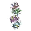
Pseudomonas aeruginosa (bacteria)
Method
X-RAY DIFFRACTION / MOLECULAR REPLACEMENT / Resolution: 2.261 Å
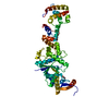
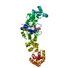
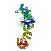
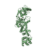
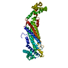
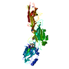
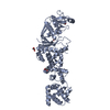
A: Exoenzyme T B: CesT family type III secretion system chaperone C: CesT family type III secretion system chaperone D: Exoenzyme T E: CesT family type III secretion system chaperone F: CesT family type III secretion system chaperone hetero molecules
In the structure databanks used in Yorodumi, some data are registered as the other names, "COVID-19 virus" and "2019-nCoV". Here are the details of the virus and the list of structure data.
Jan 31, 2019. EMDB accession codes are about to change! (news from PDBe EMDB page)
EMDB accession codes are about to change! (news from PDBe EMDB page)
The allocation of 4 digits for EMDB accession codes will soon come to an end. Whilst these codes will remain in use, new EMDB accession codes will include an additional digit and will expand incrementally as the available range of codes is exhausted. The current 4-digit format prefixed with “EMD-” (i.e. EMD-XXXX) will advance to a 5-digit format (i.e. EMD-XXXXX), and so on. It is currently estimated that the 4-digit codes will be depleted around Spring 2019, at which point the 5-digit format will come into force.
The EM Navigator/Yorodumi systems omit the EMD- prefix.
Related info.:Q: What is EMD? / ID/Accession-code notation in Yorodumi/EM Navigator
Yorodumi is a browser for structure data from EMDB, PDB, SASBDB, etc.
This page is also the successor to EM Navigator detail page, and also detail information page/front-end page for Omokage search.
The word "yorodu" (or yorozu) is an old Japanese word meaning "ten thousand". "mi" (miru) is to see.
Related info.:EMDB / PDB / SASBDB / Comparison of 3 databanks / Yorodumi Search / Aug 31, 2016. New EM Navigator & Yorodumi / Yorodumi Papers / Jmol/JSmol / Function and homology information / Changes in new EM Navigator and Yorodumi
 Movie
Movie Controller

 Yorodumi
 Open data
 Basic information
 Components
 Keywords
 Function and homology information
 Pseudomonas aeruginosa (bacteria)
 X-RAY DIFFRACTION /
  Authors
 India, 1items
  Citation
 Structure visualization
 Downloads & links
 Other downloads
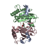


 PDBj
PDBj
 Assembly
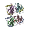

 Mass: 92.094 Da / Num. of mol.: 2 / Source method: obtained synthetically / Formula: C3H8O3
 Mass: 18.015 Da / Num. of mol.: 155 / Source method: isolated from a natural source / Formula: H2O
 Sample preparation
 Processing