 Movie
Movie Controller

[English] 日本語
 Yorodumi
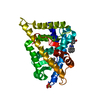
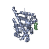
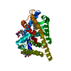
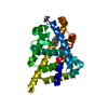
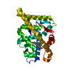
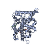
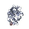
- PDB-1t79: Crystal structure of the androgen receptor ligand binding domain ... -
+ Open data

- Basic information
| Entry | Database: PDB / ID: 1t79 | ||||||
|---|---|---|---|---|---|---|---|
| Title | Crystal structure of the androgen receptor ligand binding domain in complex with a FxxLW motif | ||||||
 Components Components |
| ||||||
 Keywords Keywords | HORMONE/GROWTH FACTOR RECEPTOR /  nuclear receptor / transcription factor / ligand binding domain / AF-2 / androgen / testosterone / DHT / alpha-helical sandwich / HORMONE-GROWTH FACTOR RECEPTOR COMPLEX nuclear receptor / transcription factor / ligand binding domain / AF-2 / androgen / testosterone / DHT / alpha-helical sandwich / HORMONE-GROWTH FACTOR RECEPTOR COMPLEX | ||||||
| Function / homology |  Function and homology information Function and homology informationnegative regulation of integrin biosynthetic process / androgen binding / intracellular receptor signaling pathway / androgen receptor signaling pathway / estrogen response element binding / regulation of protein localization to plasma membrane / intracellular steroid hormone receptor signaling pathway / positive regulation of phosphorylation / steroid binding / negative regulation of extrinsic apoptotic signaling pathway ...negative regulation of integrin biosynthetic process / androgen binding / intracellular receptor signaling pathway / androgen receptor signaling pathway / estrogen response element binding / regulation of protein localization to plasma membrane / intracellular steroid hormone receptor signaling pathway / positive regulation of phosphorylation / steroid binding / negative regulation of extrinsic apoptotic signaling pathway / beta-catenin binding / male gonad development / nuclear receptor activity / positive regulation of NF-kappaB transcription factor activity / transcription cis-regulatory region binding / DNA-binding transcription factor activity / positive regulation of cell population proliferation / chromatin / positive regulation of gene expression / positive regulation of transcription by RNA polymerase II / zinc ion binding / nucleoplasm / nucleus / cytoplasmSimilarity search - Function | ||||||
| Biological species |  Pan troglodytes (chimpanzee) Pan troglodytes (chimpanzee) | ||||||
| Method | X-RAY DIFFRACTION / SYNCHROTRON / MOLECULAR REPLACEMENT / Resolution: 1.8 Å | ||||||
 Authors Authors | Hur, E. / Pfaff, S.J. / Payne, E.S. / Gron, H. / Buehrer, B.M. / Fletterick, R.J. | ||||||
 Citation Citation | Journal: Plos Biol. / Year: 2004 Title: Recognition and accommodation at the androgen receptor coactivator binding interface. Authors: Hur, E. / Pfaff, S.J. / Payne, E.S. / Gron, H. / Buehrer, B.M. / Fletterick, R.J. | ||||||
| History |
|
- Structure visualization
| Structure viewer | Molecule: MolmilJmol/JSmol |
|---|
- Downloads & links
-Download
| PDBx/mmCIF format | 1t79.cif.gz | 69.2 KB | Display | PDBx/mmCIF format |
|---|---|---|---|---|
| PDB format | pdb1t79.ent.gz | 50.5 KB | Display | PDB format |
| PDBx/mmJSON format | 1t79.json.gz | Tree view | PDBx/mmJSON format | |
| Others |  Other downloads Other downloads |
-Validation report
| Arichive directory | https://data.pdbj.org/pub/pdb/validation_reports/t7/1t79ftp://data.pdbj.org/pub/pdb/validation_reports/t7/1t79 | HTTPS FTP |
|---|
-Related structure data
| Related structure data |  1t73C  1t74C  1t76C  1t7fC 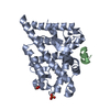 1t7mC  1t7rC  1t7tC C: citing same article ( |
|---|---|
| Similar structure data |


-Links
 PDBj
PDBj
- Assembly
| Deposited unit | 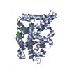
| ||||||||
|---|---|---|---|---|---|---|---|---|---|
| 1 |
| ||||||||
| Unit cell |
|
-Components
| #1: Protein | / Dihydrotestosterone receptor Mass: 30877.957 Da / Num. of mol.: 1 / Fragment: ligand binding domain Source method: isolated from a genetically manipulated source Source: (gene. exp.) Pan troglodytes (chimpanzee) / Gene: AR, NR3C4 / Production host:  Escherichia coli (E. coli) / References: UniProt: O97775 Escherichia coli (E. coli) / References: UniProt: O97775 |
|---|---|
| #2: Protein/peptide | Mass: 2123.413 Da / Num. of mol.: 1 / Source method: obtained synthetically / Details: Derived from phage display selections |
| #3: Chemical | ChemComp-DHT / Dihydrotestosterone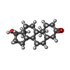  Mass: 290.440 Da / Num. of mol.: 1 / Source method: obtained synthetically / Formula: C19H30O2 / Comment: hormone*YM Mass: 290.440 Da / Num. of mol.: 1 / Source method: obtained synthetically / Formula: C19H30O2 / Comment: hormone*YM |
| #4: Water | ChemComp-HOH / Water Mass: 18.015 Da / Num. of mol.: 145 / Source method: isolated from a natural source / Formula: H2O Mass: 18.015 Da / Num. of mol.: 145 / Source method: isolated from a natural source / Formula: H2O |
-Experimental details
-Experiment
| Experiment | Method: X-RAY DIFFRACTION / Number of used crystals: 1 |
|---|
- Sample preparation
| Crystal | Density Matthews: 1.9 Å3/Da / Density % sol: 34.8 % |
|---|---|
| Crystal grow | Temperature: 293 K / Method: vapor diffusion, hanging drop / pH: 7.4 Details: sodium citrate, TRIS, HEPES, lithium sulfate, glycerol, TCEP, n-octyl glucoside, 5-alpha dihydrotestosterone, pH 7.4, VAPOR DIFFUSION, HANGING DROP, temperature 293K |
-Data collection
| Diffraction | Mean temperature: 100 K |
|---|---|
| Diffraction source | Source: SYNCHROTRON / Site: ALS  / Beamline: 8.3.1 / Wavelength: 1.12713 Å / Beamline: 8.3.1 / Wavelength: 1.12713 Å |
| Detector | Type: ADSC QUANTUM 315 / Detector: CCD / Date: May 31, 2003 |
| Radiation | Monochromator: Double Crystal Si(111) / Protocol: SINGLE WAVELENGTH / Monochromatic (M) / Laue (L): M / Scattering type: x-ray |
| Radiation wavelength | Wavelength: 1.12713 Å / Relative weight: 1 |
| Reflection | Resolution: 1.8→50 Å / Num. all: 23860 / Num. obs: 23860 / % possible obs: 99 % / Observed criterion σ(I): -3 / Biso Wilson estimate: 22.1 Å2 / Rsym value: 0.084 / Net I/σ(I): 19.1 |
| Reflection shell | Resolution: 1.8→1.83 Å / Mean I/σ(I) obs: 2.2 / Rsym value: 0.596 / % possible all: 89.6 |
- Processing
| Software |
| ||||||||||||||||||||||||||||||||||||
|---|---|---|---|---|---|---|---|---|---|---|---|---|---|---|---|---|---|---|---|---|---|---|---|---|---|---|---|---|---|---|---|---|---|---|---|---|---|
| Refinement | Method to determine structure: MOLECULAR REPLACEMENT / Resolution: 1.8→20 Å / Rfactor Rfree error: 0.007 / Data cutoff high absF: 1217201.88 / Data cutoff low absF: 0 / Isotropic thermal model: RESTRAINED / Cross valid method: THROUGHOUT / σ(F): 0 / Stereochemistry target values: Engh & Huber
| ||||||||||||||||||||||||||||||||||||
| Solvent computation | Solvent model: FLAT MODEL / Bsol: 51.5675 Å2 / ksol: 0.379274 e/Å3 | ||||||||||||||||||||||||||||||||||||
| Displacement parameters | Biso mean: 28.6 Å2
| ||||||||||||||||||||||||||||||||||||
| Refine analyze |
| ||||||||||||||||||||||||||||||||||||
| Refinement step | Cycle: LAST / Resolution: 1.8→20 Å
| ||||||||||||||||||||||||||||||||||||
| Refine LS restraints |
| ||||||||||||||||||||||||||||||||||||
| LS refinement shell | Resolution: 1.8→1.91 Å / Rfactor Rfree error: 0.024 / Total num. of bins used: 6
| ||||||||||||||||||||||||||||||||||||
| Xplor file |
|
