 Movie
Movie Controller

[English] 日本語
 Yorodumi
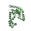
- PDB-1t3h: X-ray Structure of Dephospho-CoA Kinase from E. coli Norteast Str... -
+ Open data

- Basic information
| Entry | Database: PDB / ID: 1t3h | ||||||
|---|---|---|---|---|---|---|---|
| Title | X-ray Structure of Dephospho-CoA Kinase from E. coli Norteast Structural Genomics Consortium Target ER57 | ||||||
 Components Components | Dephospho-CoA kinase | ||||||
 Keywords Keywords | TRANSFERASE / structural genomics / PSI / Protein Structure Initiative / Northeast Structural Genomics Consortium / NESG | ||||||
| Function / homology |  Function and homology informationdephospho-CoA kinase / dephospho-CoA kinase activity / coenzyme A biosynthetic process / phosphorylation / ATP binding / cytoplasm Function and homology informationdephospho-CoA kinase / dephospho-CoA kinase activity / coenzyme A biosynthetic process / phosphorylation / ATP binding / cytoplasmSimilarity search - Function | ||||||
| Biological species |  Escherichia coli (E. coli) Escherichia coli (E. coli) | ||||||
| Method | X-RAY DIFFRACTION / SYNCHROTRON / SAD / Resolution: 2.5 Å | ||||||
 Authors Authors | Kuzin, A.P. / Chen, Y. / Forouhar, F. / Edstrom, W. / Benach, J. / Vorobiev, S. / Acton, T. / Shastry, R. / Ma, L.-C. / Xia, R. ...Kuzin, A.P. / Chen, Y. / Forouhar, F. / Edstrom, W. / Benach, J. / Vorobiev, S. / Acton, T. / Shastry, R. / Ma, L.-C. / Xia, R. / Montelione, G. / Tong, L. / Hunt, J. / Northeast Structural Genomics Consortium (NESG) | ||||||
 Citation Citation | Journal: TO BE PUBLISHED Title: X-ray Structure of Dephospho-CoA Kinase from E. coli Norteast Structural Genomics Consortium Target ER57 Authors: Kuzin, A.P. / Chen, Y. / Forouhar, F. / Edstrom, W. / Benach, J. / Vorobiev, S. / Acton, T. / Shastry, R. / Ma, L.-C. / Xia, R. / Montelione, G. / Tong, L. / Hunt, J. | ||||||
| History |
|
- Structure visualization
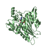
| Structure viewer | Molecule: MolmilJmol/JSmol |
|---|
- Downloads & links
-Download
| PDBx/mmCIF format | 1t3h.cif.gz | 125.2 KB | Display | PDBx/mmCIF format |
|---|---|---|---|---|
| PDB format | pdb1t3h.ent.gz | 103.4 KB | Display | PDB format |
| PDBx/mmJSON format | 1t3h.json.gz | Tree view | PDBx/mmJSON format | |
| Others |  Other downloads Other downloads |
-Validation report
| Arichive directory | https://data.pdbj.org/pub/pdb/validation_reports/t3/1t3hftp://data.pdbj.org/pub/pdb/validation_reports/t3/1t3h | HTTPS FTP |
|---|
-Related structure data
| Similar structure data | |
|---|---|
| Other databases |


-Links
 PDBj
PDBj- Assembly
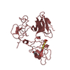
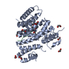
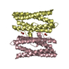
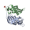
| Deposited unit | 
| ||||||||
|---|---|---|---|---|---|---|---|---|---|
| 1 | 
| ||||||||
| 2 | 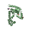
| ||||||||
| 3 | 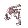
| ||||||||
| Unit cell |
|
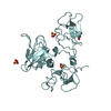
-Components
| #1: Protein | / Dephosphocoenzyme A kinase Mass: 23859.500 Da / Num. of mol.: 3 Source method: isolated from a genetically manipulated source Source: (gene. exp.) Escherichia coli (E. coli) / Production host: Escherichia coli (E. coli) / References: UniProt: P0A6I9, dephospho-CoA kinase#2: Chemical | Sulfate  Mass: 96.063 Da / Num. of mol.: 2 / Source method: obtained synthetically / Formula: SO4 Mass: 96.063 Da / Num. of mol.: 2 / Source method: obtained synthetically / Formula: SO4#3: Water | ChemComp-HOH / | Water Mass: 18.015 Da / Num. of mol.: 143 / Source method: isolated from a natural source / Formula: H2O Mass: 18.015 Da / Num. of mol.: 143 / Source method: isolated from a natural source / Formula: H2O |
|---|
-Experimental details
-Experiment
| Experiment | Method: X-RAY DIFFRACTION / Number of used crystals: 1 |
|---|
- Sample preparation
| Crystal | Density Matthews: 2.5 Å3/Da / Density % sol: 50 % |
|---|---|
| Crystal grow | Temperature: 277 K / Method: vapor diffusion, hanging drop / pH: 5.8 Details: 100 mM sodium acetate, 160 mM ammonium sulfate, 22% PEG 3350, pH 5.8, VAPOR DIFFUSION, HANGING DROP, temperature 277K |
-Data collection
| Diffraction | Mean temperature: 100 K |
|---|---|
| Diffraction source | Source: SYNCHROTRON / Site: NSLS  / Beamline: X4A / Wavelength: 0.97932 Å / Beamline: X4A / Wavelength: 0.97932 Å |
| Detector | Type: ADSC QUANTUM 4 / Detector: CCD / Date: Feb 2, 2004 |
| Radiation | Protocol: SINGLE WAVELENGTH / Monochromatic (M) / Laue (L): M / Scattering type: x-ray |
| Radiation wavelength | Wavelength: 0.97932 Å / Relative weight: 1 |
| Reflection | Resolution: 2.5→20 Å / Num. obs: 45074 / Observed criterion σ(I): -3 / Redundancy: 3.2 % / Biso Wilson estimate: 27.8 Å2 / Rmerge(I) obs: 0.081 / Rsym value: 0.081 / Net I/σ(I): 14.2 |
- Processing
| Software |
| ||||||||||||||||||||||||||||||||||||||||||||||||||||||||||||
|---|---|---|---|---|---|---|---|---|---|---|---|---|---|---|---|---|---|---|---|---|---|---|---|---|---|---|---|---|---|---|---|---|---|---|---|---|---|---|---|---|---|---|---|---|---|---|---|---|---|---|---|---|---|---|---|---|---|---|---|---|---|
| Refinement | Method to determine structure: SAD / Resolution: 2.5→19.53 Å / Rfactor Rfree error: 0.006 / Data cutoff high absF: 228923.74 / Data cutoff low absF: 0 / Isotropic thermal model: RESTRAINED / Cross valid method: THROUGHOUT / σ(F): 2 / Stereochemistry target values: Engh & Huber
| ||||||||||||||||||||||||||||||||||||||||||||||||||||||||||||
| Solvent computation | Solvent model: FLAT MODEL / Bsol: 18.62 Å2 / ksol: 0.287882 e/Å3 | ||||||||||||||||||||||||||||||||||||||||||||||||||||||||||||
| Displacement parameters | Biso mean: 33.6 Å2
| ||||||||||||||||||||||||||||||||||||||||||||||||||||||||||||
| Refine analyze |
| ||||||||||||||||||||||||||||||||||||||||||||||||||||||||||||
| Refinement step | Cycle: LAST / Resolution: 2.5→19.53 Å
| ||||||||||||||||||||||||||||||||||||||||||||||||||||||||||||
| Refine LS restraints |
| ||||||||||||||||||||||||||||||||||||||||||||||||||||||||||||
| LS refinement shell | Resolution: 2.5→2.66 Å / Rfactor Rfree error: 0.018 / Total num. of bins used: 6
| ||||||||||||||||||||||||||||||||||||||||||||||||||||||||||||
| Xplor file |
|
