 Movie
Movie Controller

[English] 日本語
 Yorodumi
- PDB-1siz: Crystal structure of the [Fe3S4]-ferredoxin from the hyperthermop... -
+ Open data

- Basic information
| Entry | Database: PDB / ID: 1siz | ||||||
|---|---|---|---|---|---|---|---|
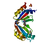
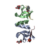
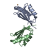
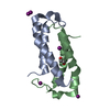
| Title | Crystal structure of the [Fe3S4]-ferredoxin from the hyperthermophilic archaeon Pyrococcus furiosus | ||||||
 Components Components | Ferredoxin | ||||||
 Keywords Keywords | ELECTRON TRANSPORT / thermostability / iron-sulfur clusters / dimer | ||||||
| Function / homology |  Function and homology information Function and homology information3 iron, 4 sulfur cluster binding / 4 iron, 4 sulfur cluster binding / electron transfer activity / iron ion binding Similarity search - Function | ||||||
| Biological species |   Pyrococcus furiosus (archaea) Pyrococcus furiosus (archaea) | ||||||
| Method |  X-RAY DIFFRACTION / SYNCHROTRON / MOLECULAR REPLACEMENT / Resolution: 2.25 Å X-RAY DIFFRACTION / SYNCHROTRON / MOLECULAR REPLACEMENT / Resolution: 2.25 Å | ||||||
 Authors Authors | Nielsen, M.S. / Harris, P. / Christensen, H.E.M. | ||||||
 Citation Citation | Journal: Biochemistry / Year: 2004 Title: The 1.5 A Resolution Crystal Structure of [Fe3S4]-Ferredoxin from the Hyperthermophilic Archaeon Pyrococcus furiosus Authors: Nielsen, M.S. / Harris, P. / Ooi, B.L. / Christensen, H.E.M. | ||||||
| History |
|
- Structure visualization
| Structure viewer | Molecule: MolmilJmol/JSmol |
|---|
- Downloads & links
-Download
| PDBx/mmCIF format | 1siz.cif.gz | 37.9 KB | Display | PDBx/mmCIF format |
|---|---|---|---|---|
| PDB format | pdb1siz.ent.gz | 26.5 KB | Display | PDB format |
| PDBx/mmJSON format | 1siz.json.gz | Tree view | PDBx/mmJSON format | |
| Others |  Other downloads Other downloads |
-Validation report
| Arichive directory | https://data.pdbj.org/pub/pdb/validation_reports/si/1sizftp://data.pdbj.org/pub/pdb/validation_reports/si/1siz | HTTPS FTP |
|---|
-Related structure data
| Related structure data |  1sj1C  1vjwS C: citing same article ( S: Starting model for refinement |
|---|---|
| Similar structure data |


-Links
 PDBj
PDBj


- Assembly
| Deposited unit | 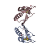
| ||||||||
|---|---|---|---|---|---|---|---|---|---|
| 1 |
| ||||||||
| Unit cell |
| ||||||||
| Details | The biological assembly is either the dimer in the asymmetric unit or a monomer |
-Components
| #1: Protein | Mass: 7171.001 Da / Num. of mol.: 2 Source method: isolated from a genetically manipulated source Source: (gene. exp.) Pyrococcus furiosus (archaea) / Gene: FDXA, PF1909 / Plasmid: pET3a / Production host:  #2: Chemical | ChemComp-3CO /   Mass: 58.933 Da / Num. of mol.: 4 / Source method: obtained synthetically / Formula: Co Mass: 58.933 Da / Num. of mol.: 4 / Source method: obtained synthetically / Formula: Co#3: Chemical |   Mass: 295.795 Da / Num. of mol.: 2 / Source method: obtained synthetically / Formula: Fe3S4 Mass: 295.795 Da / Num. of mol.: 2 / Source method: obtained synthetically / Formula: Fe3S4#4: Water | ChemComp-HOH / |  Mass: 18.015 Da / Num. of mol.: 46 / Source method: isolated from a natural source / Formula: H2O Mass: 18.015 Da / Num. of mol.: 46 / Source method: isolated from a natural source / Formula: H2OHas protein modification | Y | |
|---|
-Experimental details
-Experiment
| Experiment | Method: X-RAY DIFFRACTION / Number of used crystals: 1 |
|---|
- Sample preparation
| Crystal | Density Matthews: 2.33 Å3/Da / Density % sol: 47.18 % |
|---|---|
| Crystal grow | Temperature: 277 K / Method: vapor diffusion, hanging drop / pH: 7 Details: PEG 400, hexammine cobalt(III)-ions, HEPES, pH 7.0, VAPOR DIFFUSION, HANGING DROP, temperature 277K |
-Data collection
| Diffraction | Mean temperature: 100 K |
|---|---|
| Diffraction source | Source: SYNCHROTRON / Site: ESRF  / Beamline: ID29 / Wavelength: 0.9999 Å / Beamline: ID29 / Wavelength: 0.9999 Å |
| Detector | Type: ADSC QUANTUM 4 / Detector: CCD / Date: Apr 17, 2003 |
| Radiation | Monochromator: Silicium / Protocol: SINGLE WAVELENGTH / Monochromatic (M) / Laue (L): M / Scattering type: x-ray |
| Radiation wavelength | Wavelength: 0.9999 Å / Relative weight: 1 |
| Reflection | Resolution: 2.25→35 Å / Num. obs: 6508 / % possible obs: 94.8 % / Observed criterion σ(F): 0 / Observed criterion σ(I): 0 / Redundancy: 3.4 % / Biso Wilson estimate: 28.06 Å2 / Rmerge(I) obs: 0.1 / Net I/σ(I): 4.8 |
| Reflection shell | Resolution: 2.25→2.37 Å / Rmerge(I) obs: 0.22 / % possible all: 81.5 |
- Processing
| Software |
| ||||||||||||||||||||
|---|---|---|---|---|---|---|---|---|---|---|---|---|---|---|---|---|---|---|---|---|---|
| Refinement | Method to determine structure: MOLECULAR REPLACEMENT Starting model: pdb entry 1VJW Resolution: 2.25→35 Å / Cross valid method: THROUGHOUT / σ(F): 0 / Stereochemistry target values: Engh & Huber
| ||||||||||||||||||||
| Refinement step | Cycle: LAST / Resolution: 2.25→35 Å
| ||||||||||||||||||||
| Refine LS restraints |
|
