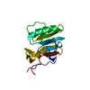
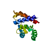
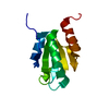
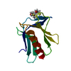
 Movie
Movie Controller


+ Open data

- Basic information
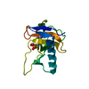
| Entry | Database: PDB / ID: 1rtu | ||||||
|---|---|---|---|---|---|---|---|
| Title | USTILAGO SPHAEROGENA RIBONUCLEASE U2 | ||||||
 Components Components | RIBONUCLEASE U2 | ||||||
 Keywords Keywords | HYDROLASE / ENDORIBONUCLEASE / BETA-ISOMERIZED ASPARTATE | ||||||
| Function / homology |  Function and homology information Function and homology informationribonuclease U2 / ribonuclease U2 activity / lyase activity / RNA binding / metal ion binding Similarity search - Function | ||||||
| Biological species |  Ustilago sphaerogena (fungus) Ustilago sphaerogena (fungus) | ||||||
| Method |  X-RAY DIFFRACTION / SYNCHROTRON / MOLECULAR REPLACEMENT / Resolution: 1.8 Å X-RAY DIFFRACTION / SYNCHROTRON / MOLECULAR REPLACEMENT / Resolution: 1.8 Å | ||||||
 Authors Authors | Noguchi, S. / Satow, Y. / Uchida, T. / Sasaki, C. / Matsuzaki, T. | ||||||
 Citation Citation | Journal: Biochemistry / Year: 1995 Title: Crystal structure of Ustilago sphaerogena ribonuclease U2 at 1.8 A resolution. Authors: Noguchi, S. / Satow, Y. / Uchida, T. / Sasaki, C. / Matsuzaki, T. | ||||||
| History |
|
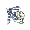
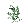
- Structure visualization
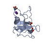
| Structure viewer | Molecule: MolmilJmol/JSmol |
|---|
- Downloads & links
-Download
| PDBx/mmCIF format | 1rtu.cif.gz | 38.2 KB | Display | PDBx/mmCIF format |
|---|---|---|---|---|
| PDB format | pdb1rtu.ent.gz | 25.2 KB | Display | PDB format |
| PDBx/mmJSON format | 1rtu.json.gz | Tree view | PDBx/mmJSON format | |
| Others |  Other downloads Other downloads |
-Validation report
| Arichive directory | https://data.pdbj.org/pub/pdb/validation_reports/rt/1rtuftp://data.pdbj.org/pub/pdb/validation_reports/rt/1rtu | HTTPS FTP |
|---|
-Related structure data
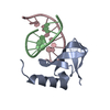
| Related structure data | 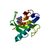 1rntS S: Starting model for refinement |
|---|---|
| Similar structure data |

-Links
 PDBj
PDBj- Assembly
| Deposited unit | 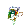
| ||||||||
|---|---|---|---|---|---|---|---|---|---|
| 1 |
| ||||||||
| Unit cell |
|
-Components
| #1: Protein | Mass: 12392.090 Da / Num. of mol.: 1 / Source method: isolated from a natural source / Source: (natural) Ustilago sphaerogena (fungus) / References: UniProt: P00654 | ||
|---|---|---|---|
| #2: Chemical | ChemComp-SO4 /   Mass: 96.063 Da / Num. of mol.: 1 / Source method: obtained synthetically / Formula: SO4 Mass: 96.063 Da / Num. of mol.: 1 / Source method: obtained synthetically / Formula: SO4 | ||
| #3: Water | ChemComp-HOH /  Mass: 18.015 Da / Num. of mol.: 141 / Source method: isolated from a natural source / Formula: H2O Mass: 18.015 Da / Num. of mol.: 141 / Source method: isolated from a natural source / Formula: H2O | ||
| Compound details | ASP 45 IS ISOMERIZED| Has protein modification | Y | |
-Experimental details
-Experiment
| Experiment | Method: X-RAY DIFFRACTION / Number of used crystals: 1 |
|---|
- Sample preparation
| Crystal | Density Matthews: 2.13 Å3/Da / Density % sol: 42.4 % | ||||||||||||||||||||||||||||||
|---|---|---|---|---|---|---|---|---|---|---|---|---|---|---|---|---|---|---|---|---|---|---|---|---|---|---|---|---|---|---|---|
| Crystal grow | Temperature: 293 K / Method: vapor diffusion, hanging drop / pH: 4.5 Details: CRYSTALS WERE PREPARED BY HANGING-DROP VAPOUR-DIFFUSION METHOD AT 293 KELVIN FROM A 20 MG/ML PROTEIN SOLUTION CONTAINING 15 MG/ML 2'-DEOXY 2'-FLUORO ADENYLYL-3',5'-CYTIDINE, 0.4M AMMONIUM ...Details: CRYSTALS WERE PREPARED BY HANGING-DROP VAPOUR-DIFFUSION METHOD AT 293 KELVIN FROM A 20 MG/ML PROTEIN SOLUTION CONTAINING 15 MG/ML 2'-DEOXY 2'-FLUORO ADENYLYL-3',5'-CYTIDINE, 0.4M AMMONIUM SULFATE, EQUILIBRATED AGAINST A RESERVOIR OF 0.9M AMMONIUM SULFATE CONTAINING 0.1M ACETATE BUFFER (PH 4.5)., vapor diffusion - hanging drop | ||||||||||||||||||||||||||||||
| Crystal grow | *PLUS Temperature: 20 ℃ / Method: vapor diffusion | ||||||||||||||||||||||||||||||
| Components of the solutions | *PLUS
|
-Data collection
| Diffraction | Mean temperature: 284 K |
|---|---|
| Diffraction source | Source: SYNCHROTRON / Site: Photon Factory  / Type: PHOTON FACTORY / Wavelength: 0.9 / Type: PHOTON FACTORY / Wavelength: 0.9 |
| Detector | Type: FUJI / Detector: IMAGE PLATE / Date: May 22, 1990 / Details: DOUBLE FOCUSSING MIRROR |
| Radiation | Monochromator: SI(111) / Monochromatic (M) / Laue (L): M / Scattering type: x-ray |
| Radiation wavelength | Wavelength: 0.9 Å / Relative weight: 1 |
| Reflection | Resolution: 1.8→12 Å / Num. obs: 9903 / % possible obs: 96.1 % / Observed criterion σ(I): 1 / Redundancy: 4 % / Rmerge(I) obs: 0.0342 |
| Reflection shell | Resolution: 1.8→1.87 Å / Redundancy: 3 % / Rmerge(I) obs: 0.077 / % possible all: 86.4 |
| Reflection | *PLUS Highest resolution: 1.8 Å / Rmerge(I) obs: 0.044 |
| Reflection shell | *PLUS |
- Processing
| Software |
| ||||||||||||||||||||||||||||||||||||||||||||||||||||||||||||||||||||||||||||||||||||
|---|---|---|---|---|---|---|---|---|---|---|---|---|---|---|---|---|---|---|---|---|---|---|---|---|---|---|---|---|---|---|---|---|---|---|---|---|---|---|---|---|---|---|---|---|---|---|---|---|---|---|---|---|---|---|---|---|---|---|---|---|---|---|---|---|---|---|---|---|---|---|---|---|---|---|---|---|---|---|---|---|---|---|---|---|---|
| Refinement | Method to determine structure: MOLECULAR REPLACEMENT Starting model: RNASE T1 (PDB ENTRY 1RNT) Resolution: 1.8→8 Å / σ(F): 0
| ||||||||||||||||||||||||||||||||||||||||||||||||||||||||||||||||||||||||||||||||||||
| Displacement parameters | Biso mean: 13.4 Å2 | ||||||||||||||||||||||||||||||||||||||||||||||||||||||||||||||||||||||||||||||||||||
| Refine analyze | Luzzati coordinate error obs: 0.13 Å | ||||||||||||||||||||||||||||||||||||||||||||||||||||||||||||||||||||||||||||||||||||
| Refinement step | Cycle: LAST / Resolution: 1.8→8 Å
| ||||||||||||||||||||||||||||||||||||||||||||||||||||||||||||||||||||||||||||||||||||
| Refine LS restraints |
| ||||||||||||||||||||||||||||||||||||||||||||||||||||||||||||||||||||||||||||||||||||
| Software | *PLUS Name: PROLSQ / Classification: refinement | ||||||||||||||||||||||||||||||||||||||||||||||||||||||||||||||||||||||||||||||||||||
| Refinement | *PLUS % reflection Rfree: 10 % / Rfactor Rfree: 0.178 | ||||||||||||||||||||||||||||||||||||||||||||||||||||||||||||||||||||||||||||||||||||
| Solvent computation | *PLUS | ||||||||||||||||||||||||||||||||||||||||||||||||||||||||||||||||||||||||||||||||||||
| Displacement parameters | *PLUS |
