 Movie
Movie Controller

[English] 日本語
 Yorodumi
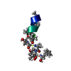
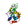
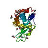
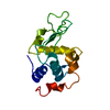
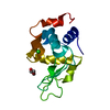
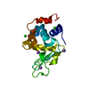
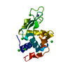
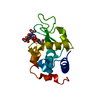
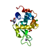
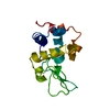
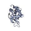
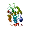
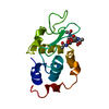
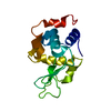
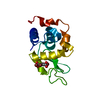
- PDB-1qqy: X-RAY CRYSTAL STRUCTURE ANALYSIS OF CANINE MILK LYSOZYME (APO-TYPE) -
+ Open data

- Basic information
| Entry | Database: PDB / ID: 1qqy | ||||||
|---|---|---|---|---|---|---|---|
| Title | X-RAY CRYSTAL STRUCTURE ANALYSIS OF CANINE MILK LYSOZYME (APO-TYPE) | ||||||
 Components Components | LYSOZYME C | ||||||
 Keywords Keywords | HYDROLASE / APO-TYPE PROTEIN / CALCIUM BINDING LYSOZYME / ENZYME | ||||||
| Function / homology |  Function and homology information Function and homology informationlysozyme / lysozyme activity / killing of cells of another organism / defense response to bacterium / metal ion binding Similarity search - Function | ||||||
| Biological species |  | ||||||
| Method |  X-RAY DIFFRACTION / SYNCHROTRON / MOLECULAR REPLACEMENT / Resolution: 1.85 Å X-RAY DIFFRACTION / SYNCHROTRON / MOLECULAR REPLACEMENT / Resolution: 1.85 Å | ||||||
 Authors Authors | Koshiba, T. / Yao, M. / Tanaka, I. / Nitta, K. | ||||||
 Citation Citation | Journal: Biochemistry / Year: 2000 Title: Structure and thermodynamics of the extraordinarily stable molten globule state of canine milk lysozyme. Authors: Koshiba, T. / Yao, M. / Kobashigawa, Y. / Demura, M. / Nakagawa, A. / Tanaka, I. / Kuwajima, K. / Nitta, K. #1: Journal: Protein Eng. / Year: 1999Title: Expression of a synthetic gene encoding canine milk lysozyme in Escherichia coli and characterization of the expressed protein Authors: Koshiba, T. / Hayashi, T. / Miwako, I. / Kumagai, I. / Ikura, T. / Kawano, K. / Nitta, K. / Kuwajima, K. | ||||||
| History |
|
- Structure visualization
| Structure viewer | Molecule: MolmilJmol/JSmol |
|---|
- Downloads & links
-Download
| PDBx/mmCIF format | 1qqy.cif.gz | 38.9 KB | Display | PDBx/mmCIF format |
|---|---|---|---|---|
| PDB format | pdb1qqy.ent.gz | 26.7 KB | Display | PDB format |
| PDBx/mmJSON format | 1qqy.json.gz | Tree view | PDBx/mmJSON format | |
| Others |  Other downloads Other downloads |
-Validation report
| Summary document | 1qqy_validation.pdf.gz | 359 KB | Display | wwPDB validaton report |
|---|---|---|---|---|
| Full document | 1qqy_full_validation.pdf.gz | 362.2 KB | Display | |
| Data in XML | 1qqy_validation.xml.gz | 4 KB | Display | |
| Data in CIF | 1qqy_validation.cif.gz | 6 KB | Display | |
| Arichive directory | https://data.pdbj.org/pub/pdb/validation_reports/qq/1qqyftp://data.pdbj.org/pub/pdb/validation_reports/qq/1qqy | HTTPS FTP |
-Related structure data
| Related structure data |  1eqlS S: Starting model for refinement |
|---|---|
| Similar structure data |


-Links
 PDBj
PDBj- Assembly
| Deposited unit | 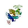
| ||||||||
|---|---|---|---|---|---|---|---|---|---|
| 1 |
| ||||||||
| Unit cell |
|
-Components
| #1: Protein | Mass: 14621.643 Da / Num. of mol.: 1 Source method: isolated from a genetically manipulated source Source: (gene. exp.)  |
|---|---|
| #2: Water | ChemComp-HOH /  Mass: 18.015 Da / Num. of mol.: 81 / Source method: isolated from a natural source / Formula: H2O Mass: 18.015 Da / Num. of mol.: 81 / Source method: isolated from a natural source / Formula: H2O |
-Experimental details
-Experiment
| Experiment | Method: X-RAY DIFFRACTION / Number of used crystals: 1 |
|---|
- Sample preparation
| Crystal | Density Matthews: 2.5 Å3/Da / Density % sol: 51.5 % | ||||||||||||||||||||||||||||||
|---|---|---|---|---|---|---|---|---|---|---|---|---|---|---|---|---|---|---|---|---|---|---|---|---|---|---|---|---|---|---|---|
| Crystal grow | Temperature: 291 K / Method: vapor diffusion, hanging drop / pH: 5.6 Details: PEG 4000, tri-sodium citrate, ammonium acetate, EDTA, pH 5.6, VAPOR DIFFUSION, HANGING DROP, temperature 291.0K | ||||||||||||||||||||||||||||||
| Crystal grow | *PLUS | ||||||||||||||||||||||||||||||
| Components of the solutions | *PLUS
|
-Data collection
| Diffraction | Mean temperature: 277 K |
|---|---|
| Diffraction source | Source: SYNCHROTRON / Site: Photon Factory  / Beamline: BL-18B / Wavelength: 1 / Beamline: BL-18B / Wavelength: 1 |
| Detector | Type: WEISSENBERG / Detector: DIFFRACTOMETER / Date: Nov 25, 1998 |
| Radiation | Protocol: SINGLE WAVELENGTH / Monochromatic (M) / Laue (L): M / Scattering type: x-ray |
| Radiation wavelength | Wavelength: 1 Å / Relative weight: 1 |
| Reflection | Resolution: 1.85→100 Å / Num. obs: 13034 / % possible obs: 94.8 % / Observed criterion σ(I): 1 / Redundancy: 15.3 % / Biso Wilson estimate: 27.6 Å2 / Rmerge(I) obs: 0.06 / Net I/σ(I): 12.9 |
| Reflection shell | Resolution: 1.85→1.89 Å / Rmerge(I) obs: 0.25 / Num. unique all: 725 / % possible all: 82.2 |
| Reflection | *PLUS Num. measured all: 199512 |
| Reflection shell | *PLUS % possible obs: 82.2 % |
- Processing
| Software |
| |||||||||||||||||||||||||
|---|---|---|---|---|---|---|---|---|---|---|---|---|---|---|---|---|---|---|---|---|---|---|---|---|---|---|
| Refinement | Method to determine structure: MOLECULAR REPLACEMENT Starting model: 1EQL Resolution: 1.85→8 Å / Rfactor Rfree error: 0.006 / Cross valid method: THROUGHOUT / σ(F): 2 / Stereochemistry target values: Engh & Huber
| |||||||||||||||||||||||||
| Solvent computation | Solvent model: THROUGHOUT / Bsol: 62.4 Å2 / ksol: 0.449 e/Å3 | |||||||||||||||||||||||||
| Displacement parameters | Biso mean: 26.2 Å2
| |||||||||||||||||||||||||
| Refine analyze |
| |||||||||||||||||||||||||
| Refinement step | Cycle: LAST / Resolution: 1.85→8 Å
| |||||||||||||||||||||||||
| Refine LS restraints |
| |||||||||||||||||||||||||
| LS refinement shell | Resolution: 1.85→1.88 Å / Rfactor Rfree error: 0.043 / Total num. of bins used: 24
| |||||||||||||||||||||||||
| Xplor file | Serial no: 1 / Param file: PROTEIN_REP.PARAM / Topol file: PROTEIN.TOP | |||||||||||||||||||||||||
| Software | *PLUS Name: 'CNS' / Classification: refinement | |||||||||||||||||||||||||
| Refine LS restraints | *PLUS Type: c_bond_d / Dev ideal: 0.007 |
