 Movie
Movie Controller


+ Open data

- Basic information
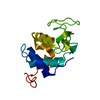
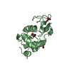
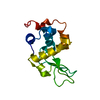
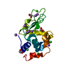
| Entry | Database: PDB / ID: 1lyy | ||||||
|---|---|---|---|---|---|---|---|
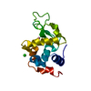
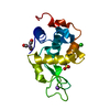
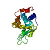
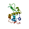
| Title | AMYLOIDOGENIC VARIANT (ASP67HIS) OF HUMAN LYSOZYME | ||||||
 Components Components | LYSOZYME | ||||||
 Keywords Keywords | HYDROLASE / ENZYME / BETA-1 / 4-GLYCAN-HYDROLASE | ||||||
| Function / homology |  Function and homology information Function and homology informationantimicrobial humoral response / Antimicrobial peptides / specific granule lumen / lysozyme / lysozyme activity / azurophil granule lumen / tertiary granule lumen / killing of cells of another organism / defense response to Gram-negative bacterium / defense response to bacterium ...antimicrobial humoral response / Antimicrobial peptides / specific granule lumen / lysozyme / lysozyme activity / azurophil granule lumen / tertiary granule lumen / killing of cells of another organism / defense response to Gram-negative bacterium / defense response to bacterium / defense response to Gram-positive bacterium / Amyloid fiber formation / inflammatory response / Neutrophil degranulation / : / extracellular exosome / extracellular region / identical protein binding Similarity search - Function | ||||||
| Biological species |  Homo sapiens (human) Homo sapiens (human) | ||||||
| Method |  X-RAY DIFFRACTION / MOLECULAR REPLACEMENT / Resolution: 1.8 Å X-RAY DIFFRACTION / MOLECULAR REPLACEMENT / Resolution: 1.8 Å | ||||||
 Authors Authors | Sunde, M. / Blake, C.C.F. | ||||||
 Citation Citation | Journal: Nature / Year: 1997 Title: Instability, unfolding and aggregation of human lysozyme variants underlying amyloid fibrillogenesis. Authors: Booth, D.R. / Sunde, M. / Bellotti, V. / Robinson, C.V. / Hutchinson, W.L. / Fraser, P.E. / Hawkins, P.N. / Dobson, C.M. / Radford, S.E. / Blake, C.C. / Pepys, M.B. | ||||||
| History |
|
- Structure visualization
| Structure viewer | Molecule: MolmilJmol/JSmol |
|---|
- Downloads & links
-Download
| PDBx/mmCIF format | 1lyy.cif.gz | 39.9 KB | Display | PDBx/mmCIF format |
|---|---|---|---|---|
| PDB format | pdb1lyy.ent.gz | 27.3 KB | Display | PDB format |
| PDBx/mmJSON format | 1lyy.json.gz | Tree view | PDBx/mmJSON format | |
| Others |  Other downloads Other downloads |
-Validation report
| Arichive directory | https://data.pdbj.org/pub/pdb/validation_reports/ly/1lyyftp://data.pdbj.org/pub/pdb/validation_reports/ly/1lyy | HTTPS FTP |
|---|
-Related structure data


-Links
 PDBj
PDBj

- Assembly
| Deposited unit | 
| ||||||||
|---|---|---|---|---|---|---|---|---|---|
| 1 |
| ||||||||
| Unit cell |
|
-Components
| #1: Protein | Mass: 14743.752 Da / Num. of mol.: 1 / Mutation: D67H Source method: isolated from a genetically manipulated source Source: (gene. exp.) Homo sapiens (human) / Description: HUMAN LYSOZYME CDNA AMPLIFIED FROM RNA / Plasmid: PBACPAK8 / Production host:   Spodoptera frugiperda (fall armyworm) / Strain (production host): SF9 / References: UniProt: P61626, lysozyme Spodoptera frugiperda (fall armyworm) / Strain (production host): SF9 / References: UniProt: P61626, lysozyme |
|---|---|
| #2: Water | ChemComp-HOH /  Mass: 18.015 Da / Num. of mol.: 106 / Source method: isolated from a natural source / Formula: H2O Mass: 18.015 Da / Num. of mol.: 106 / Source method: isolated from a natural source / Formula: H2O |
| Has protein modification | Y |
-Experimental details
-Experiment
| Experiment | Method: X-RAY DIFFRACTION / Number of used crystals: 1 |
|---|
- Sample preparation
| Crystal | Density Matthews: 2.04 Å3/Da / Density % sol: 40 % | ||||||||||||||||||||||||||||||||||||
|---|---|---|---|---|---|---|---|---|---|---|---|---|---|---|---|---|---|---|---|---|---|---|---|---|---|---|---|---|---|---|---|---|---|---|---|---|---|
| Crystal grow | pH: 4 Details: PROTEIN WAS CRYSTALLIZED FROM 0.2 M AMMONIUM SULFATE, 30% PEG 8000, pH 4.0 | ||||||||||||||||||||||||||||||||||||
| Crystal grow | *PLUS Temperature: 20 ℃ / Method: vapor diffusion / PH range low: 8 / PH range high: 7.1 | ||||||||||||||||||||||||||||||||||||
| Components of the solutions | *PLUS
|
-Data collection
| Diffraction | Mean temperature: 288 K |
|---|---|
| Diffraction source | Source: ROTATING ANODE / Type: RIGAKU / Wavelength: 1.5418 |
| Detector | Type: MARRESEARCH / Detector: IMAGE PLATE / Date: Jan 1, 1995 |
| Radiation | Monochromator: GRAPHITE(002) / Monochromatic (M) / Laue (L): M / Scattering type: x-ray |
| Radiation wavelength | Wavelength: 1.5418 Å / Relative weight: 1 |
| Reflection | Resolution: 1.75→30 Å / Num. obs: 11071 / % possible obs: 90.9 % / Observed criterion σ(I): 2 / Redundancy: 3.7 % / Rmerge(I) obs: 0.103 / Rsym value: 0.131 / Net I/σ(I): 8.5 |
| Reflection shell | Resolution: 1.75→1.81 Å / Redundancy: 2.1 % / Rmerge(I) obs: 0.198 / Mean I/σ(I) obs: 3 / Rsym value: 0.223 / % possible all: 78.3 |
| Reflection | *PLUS Num. measured all: 41333 |
| Reflection shell | *PLUS % possible obs: 78.3 % |
- Processing
| Software |
| ||||||||||||||||||||||||||||||||||||||||||||||||||||||||||||
|---|---|---|---|---|---|---|---|---|---|---|---|---|---|---|---|---|---|---|---|---|---|---|---|---|---|---|---|---|---|---|---|---|---|---|---|---|---|---|---|---|---|---|---|---|---|---|---|---|---|---|---|---|---|---|---|---|---|---|---|---|---|
| Refinement | Method to determine structure: MOLECULAR REPLACEMENT Starting model: HUMAN LYSOZYME COORDINATES FROM ARTYMUIK, P.J.AND BLAKE, C.C.F. J.MOL.BIOL. (1983) 167,693-723. Resolution: 1.8→8 Å / Isotropic thermal model: RESTRAINED Cross valid method: NOT USED AFTER INITIAL STAGES BECAUSE OF LOW NUMBER OF REFLECTIONS σ(F): 0
| ||||||||||||||||||||||||||||||||||||||||||||||||||||||||||||
| Displacement parameters | Biso mean: 14.8 Å2 | ||||||||||||||||||||||||||||||||||||||||||||||||||||||||||||
| Refinement step | Cycle: LAST / Resolution: 1.8→8 Å
| ||||||||||||||||||||||||||||||||||||||||||||||||||||||||||||
| Refine LS restraints |
| ||||||||||||||||||||||||||||||||||||||||||||||||||||||||||||
| Xplor file |
| ||||||||||||||||||||||||||||||||||||||||||||||||||||||||||||
| Software | *PLUS Name: X-PLOR / Classification: refinement | ||||||||||||||||||||||||||||||||||||||||||||||||||||||||||||
| Refinement | *PLUS | ||||||||||||||||||||||||||||||||||||||||||||||||||||||||||||
| Solvent computation | *PLUS | ||||||||||||||||||||||||||||||||||||||||||||||||||||||||||||
| Displacement parameters | *PLUS | ||||||||||||||||||||||||||||||||||||||||||||||||||||||||||||
| Refine LS restraints | *PLUS
|
