 Movie
Movie Controller

[English] 日本語
 Yorodumi
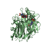
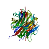
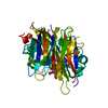
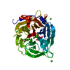
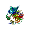
- PDB-1qlg: Crystal structure of phytase with magnesium from Bacillus amyloli... -
+ Open data

- Basic information
| Entry | Database: PDB / ID: 1qlg | ||||||
|---|---|---|---|---|---|---|---|
| Title | Crystal structure of phytase with magnesium from Bacillus amyloliquefaciens | ||||||
 Components Components | 3-PHYTASE | ||||||
 Keywords Keywords | PHOSPHOMONOESTERASE / PHYTASE / THERMOSTABLE / PHOSPHATASE / CALCIUM / MAGNESIUM | ||||||
| Function / homology |  Function and homology information Function and homology information3-phytase / inositol hexakisphosphate 3-phosphatase activity / extracellular region Similarity search - Function | ||||||
| Biological species |  | ||||||
| Method |  X-RAY DIFFRACTION / DIRECT METHODS / Resolution: 2.2 Å X-RAY DIFFRACTION / DIRECT METHODS / Resolution: 2.2 Å | ||||||
 Authors Authors | Shin, S. / Ha, N.-C. / Oh, B.-H. | ||||||
 Citation Citation | Journal: Nat.Struct.Biol. / Year: 2000 Title: Crystal Structures of a Novel, Thermostable Phytase in Partially and Fully Calcium-Loaded States Authors: Ha, N.-C. / Oh, B.-C. / Shin, S. / Kim, H.J. / Oh, T.K. / Kim, Y.O. / Choi, K.Y. / Oh, B.H. | ||||||
| History |
|
- Structure visualization
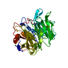
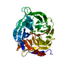
| Structure viewer | Molecule: MolmilJmol/JSmol |
|---|
- Downloads & links
-Download
| PDBx/mmCIF format | 1qlg.cif.gz | 84.1 KB | Display | PDBx/mmCIF format |
|---|---|---|---|---|
| PDB format | pdb1qlg.ent.gz | 61.1 KB | Display | PDB format |
| PDBx/mmJSON format | 1qlg.json.gz | Tree view | PDBx/mmJSON format | |
| Others |  Other downloads Other downloads |
-Validation report
| Arichive directory | https://data.pdbj.org/pub/pdb/validation_reports/ql/1qlgftp://data.pdbj.org/pub/pdb/validation_reports/ql/1qlg | HTTPS FTP |
|---|
-Related structure data
| Related structure data | 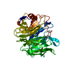 1cvmC  1pooSC 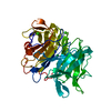 2pooC S: Starting model for refinement C: citing same article ( |
|---|---|
| Similar structure data |


-Links
 PDBj
PDBj


- Assembly
| Deposited unit | 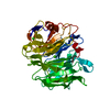
| ||||||||
|---|---|---|---|---|---|---|---|---|---|
| 1 |
| ||||||||
| Unit cell |
| ||||||||
| Details | BIOLOGICAL_UNIT: MONOMER |
-Components
| #1: Protein | Mass: 39007.801 Da / Num. of mol.: 1 Source method: isolated from a genetically manipulated source Details: CALCIUM MAGNESIUM / Source: (gene. exp.) | ||||
|---|---|---|---|---|---|
| #2: Chemical |   Mass: 40.078 Da / Num. of mol.: 3 / Source method: obtained synthetically / Formula: Ca Mass: 40.078 Da / Num. of mol.: 3 / Source method: obtained synthetically / Formula: Ca#3: Chemical |   Mass: 24.305 Da / Num. of mol.: 2 / Source method: obtained synthetically / Formula: Mg Mass: 24.305 Da / Num. of mol.: 2 / Source method: obtained synthetically / Formula: Mg#4: Water | ChemComp-HOH / |  Mass: 18.015 Da / Num. of mol.: 48 / Source method: isolated from a natural source / Formula: H2O Mass: 18.015 Da / Num. of mol.: 48 / Source method: isolated from a natural source / Formula: H2O |
-Experimental details
-Experiment
| Experiment | Method: X-RAY DIFFRACTION / Number of used crystals: 1 |
|---|
- Sample preparation
| Crystal | Density Matthews: 2.21 Å3/Da / Density % sol: 44.47 % | ||||||||||||||||||||||||
|---|---|---|---|---|---|---|---|---|---|---|---|---|---|---|---|---|---|---|---|---|---|---|---|---|---|
| Crystal grow | pH: 6.5 / Details: pH 6.50 | ||||||||||||||||||||||||
| Crystal grow | *PLUS Temperature: 277 K / Method: vapor diffusion, hanging dropDetails: drop consists of equal volume of protein and precipitant solutions | ||||||||||||||||||||||||
| Components of the solutions | *PLUS
|
-Data collection
| Diffraction | Mean temperature: 296 K |
|---|---|
| Diffraction source | Source: ROTATING ANODE / Type: MACSCIENCE / Wavelength: 1.5418 |
| Detector | Type: MACSCIENCE / Detector: IMAGE PLATE / Details: MONOCHROMETER |
| Radiation | Monochromator: GRAPHITE(002) / Protocol: SINGLE WAVELENGTH / Monochromatic (M) / Laue (L): M / Scattering type: x-ray |
| Radiation wavelength | Wavelength: 1.5418 Å / Relative weight: 1 |
| Reflection | Resolution: 2.2→40 Å / Num. obs: 17305 / % possible obs: 93 % / Observed criterion σ(I): 1 / Redundancy: 1.07 % / Rmerge(I) obs: 0.084 / Rsym value: 0.084 / Net I/σ(I): 13.5 |
| Reflection shell | Resolution: 2.2→2.28 Å / Redundancy: 1 % / Rmerge(I) obs: 0.317 / Mean I/σ(I) obs: 2.62 / Rsym value: 0.317 / % possible all: 87.2 |
| Reflection shell | *PLUS % possible obs: 87.2 % |
- Processing
| Software |
| ||||||||||||||||||||||||||||||||||||||||||||||||||||||||||||
|---|---|---|---|---|---|---|---|---|---|---|---|---|---|---|---|---|---|---|---|---|---|---|---|---|---|---|---|---|---|---|---|---|---|---|---|---|---|---|---|---|---|---|---|---|---|---|---|---|---|---|---|---|---|---|---|---|---|---|---|---|---|
| Refinement | Method to determine structure: DIRECT METHODS Starting model: 1POO Resolution: 2.2→8 Å / Cross valid method: THROUGHOUT / σ(F): 2
| ||||||||||||||||||||||||||||||||||||||||||||||||||||||||||||
| Refinement step | Cycle: LAST / Resolution: 2.2→8 Å
| ||||||||||||||||||||||||||||||||||||||||||||||||||||||||||||
| Refine LS restraints |
|
