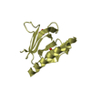
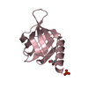
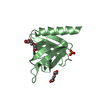
登録情報 データベース : PDB / ID : 1q7xタイトル Solution structure of the alternatively spliced PDZ2 domain (PDZ2b) of PTP-Bas (hPTP1E) PDZ2b domain of PTP-Bas (hPTP1E) キーワード / / / / 機能・相同性 分子機能 ドメイン・相同性 構成要素
/ / / / / / / / / / / / / / / / / / / / / / / / / / / / / / / / / / / / / / / / / / / / / / / / / / / / / / / / / / / / / / / / / / / / / / / / / / / / / 生物種 Homo sapiens (ヒト)手法 / データ登録者 Kachel, N. / Erdmann, K.S. / Kremer, W. / Wolff, P. / Gronwald, W. / Heumann, R. / Kalbitzer, H.R. / Structural Proteomics in Europe (SPINE) ジャーナル : J.Mol.Biol. / 年 : 2003タイトル : Structure determination and ligand interactions of the PDZ2b domain of PTP-Bas (hPTP1E): Splicing induced modulation of ligand specificity.著者 : Kachel, N. / Erdmann, K.S. / Kremer, W. / Wolff, P. / Gronwald, W. / Heumann, R. / Kalbitzer, H.R. 履歴 登録 2003年8月20日 登録サイト / 処理サイト 改定 1.0 2003年12月2日 Provider / タイプ 改定 1.1 2008年4月29日 Group 改定 1.2 2011年7月13日 Group 改定 1.3 2022年3月2日 Group / Database references / Derived calculationsカテゴリ database_2 / pdbx_nmr_software ... database_2 / pdbx_nmr_software / pdbx_struct_assembly / pdbx_struct_oper_list / struct_ref_seq_dif Item _database_2.pdbx_DOI / _database_2.pdbx_database_accession ... _database_2.pdbx_DOI / _database_2.pdbx_database_accession / _pdbx_nmr_software.name / _struct_ref_seq_dif.details 改定 1.4 2024年5月22日 Group / カテゴリ / chem_comp_bond
すべて表示 表示を減らす
 ムービー
ムービー コントローラー


 データを開く
 基本情報
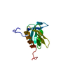
基本情報 要素
要素 キーワード
キーワード 機能・相同性情報
 Homo sapiens (ヒト)
 データ登録者
 引用
引用 構造の表示
構造の表示 ダウンロードとリンク
 その他のダウンロード
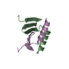


 PDBj
PDBj


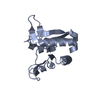
 集合体
集合体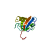

 試料調製
試料調製 解析
解析