 Movie
Movie Controller


+ Open data

- Basic information
| Entry | Database: PDB / ID: 1q67 | ||||||
|---|---|---|---|---|---|---|---|
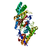
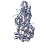
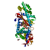
| Title | Crystal structure of Dcp1p | ||||||
 Components Components | Decapping protein involved in mRNA degradation-Dcp1p | ||||||
 Keywords Keywords | TRANSCRIPTION / beta sandwich | ||||||
| Function / homology |  Function and homology information Function and homology informationRNA decapping complex / Butyrate Response Factor 1 (BRF1) binds and destabilizes mRNA / Tristetraprolin (TTP, ZFP36) binds and destabilizes mRNA / mRNA decay by 5' to 3' exoribonuclease / deadenylation-independent decapping of nuclear-transcribed mRNA / deadenylation-dependent decapping of nuclear-transcribed mRNA / cytoplasmic side of membrane / nuclear-transcribed mRNA catabolic process, nonsense-mediated decay / P-body / enzyme activator activity ...RNA decapping complex / Butyrate Response Factor 1 (BRF1) binds and destabilizes mRNA / Tristetraprolin (TTP, ZFP36) binds and destabilizes mRNA / mRNA decay by 5' to 3' exoribonuclease / deadenylation-independent decapping of nuclear-transcribed mRNA / deadenylation-dependent decapping of nuclear-transcribed mRNA / cytoplasmic side of membrane / nuclear-transcribed mRNA catabolic process, nonsense-mediated decay / P-body / enzyme activator activity / mRNA processing / mRNA binding / nucleus / cytoplasm Similarity search - Function | ||||||
| Biological species |  | ||||||
| Method |  X-RAY DIFFRACTION / SYNCHROTRON / SAD / Resolution: 2.3 Å X-RAY DIFFRACTION / SYNCHROTRON / SAD / Resolution: 2.3 Å | ||||||
 Authors Authors | She, M. / Decker, C.J. / Liu, Y. / Chen, N. / Parker, R. / Song, H. | ||||||
 Citation Citation | Journal: Nat.Struct.Mol.Biol. / Year: 2004 Title: Crystal structure of Dcp1p and its functional implications in mRNA decapping Authors: She, M. / Decker, C.J. / Sundramurthy, K. / Liu, Y. / Chen, N. / Parker, R. / Song, H. #1: Journal: Nature / Year: 1996Title: An essential component of the decapping enzyme required for normal rates of mRNA turnover Authors: Beelman, C.A. / Stevens, A. / Caponigro, G. / LaGrandeur, T.E. / Hatfield, L. / Fortner, D.M. / Parker, R. | ||||||
| History |
|
- Structure visualization
| Structure viewer | Molecule: MolmilJmol/JSmol |
|---|
- Downloads & links
-Download
| PDBx/mmCIF format | 1q67.cif.gz | 79.5 KB | Display | PDBx/mmCIF format |
|---|---|---|---|---|
| PDB format | pdb1q67.ent.gz | 59.8 KB | Display | PDB format |
| PDBx/mmJSON format | 1q67.json.gz | Tree view | PDBx/mmJSON format | |
| Others |  Other downloads Other downloads |
-Validation report
| Arichive directory | https://data.pdbj.org/pub/pdb/validation_reports/q6/1q67ftp://data.pdbj.org/pub/pdb/validation_reports/q6/1q67 | HTTPS FTP |
|---|
-Related structure data
| Similar structure data |
|---|


-Links
 PDBj
PDBj
- Assembly
| Deposited unit | 
| ||||||||
|---|---|---|---|---|---|---|---|---|---|
| 1 |
| ||||||||
| Unit cell |
|
-Components
| #1: Protein | Mass: 26294.357 Da / Num. of mol.: 2 Source method: isolated from a genetically manipulated source Source: (gene. exp.) Gene: DCP1 / Production host:  #2: Water | ChemComp-HOH / |  Mass: 18.015 Da / Num. of mol.: 156 / Source method: isolated from a natural source / Formula: H2O Mass: 18.015 Da / Num. of mol.: 156 / Source method: isolated from a natural source / Formula: H2O |
|---|
-Experimental details
-Experiment
| Experiment | Method: X-RAY DIFFRACTION / Number of used crystals: 1 |
|---|
- Sample preparation
| Crystal | Density Matthews: 3.26 Å3/Da / Density % sol: 62.3 % | ||||||||||||||||||||||||||||||||||||||||||||||||||||||||||||||||||||||||||||||||||||
|---|---|---|---|---|---|---|---|---|---|---|---|---|---|---|---|---|---|---|---|---|---|---|---|---|---|---|---|---|---|---|---|---|---|---|---|---|---|---|---|---|---|---|---|---|---|---|---|---|---|---|---|---|---|---|---|---|---|---|---|---|---|---|---|---|---|---|---|---|---|---|---|---|---|---|---|---|---|---|---|---|---|---|---|---|---|
| Crystal grow | Details: VAPOR DIFFUSION, HANGING DROP | ||||||||||||||||||||||||||||||||||||||||||||||||||||||||||||||||||||||||||||||||||||
| Crystal grow | *PLUS pH: 7.6 / Method: vapor diffusion, hanging drop | ||||||||||||||||||||||||||||||||||||||||||||||||||||||||||||||||||||||||||||||||||||
| Components of the solutions | *PLUS
|
-Data collection
| Diffraction | Mean temperature: 100 K |
|---|---|
| Diffraction source | Source: SYNCHROTRON / Site: SPring-8  / Beamline: BL41XU / Wavelength: 0.9798 / Beamline: BL41XU / Wavelength: 0.9798 |
| Detector | Type: MARRESEARCH / Detector: CCD |
| Radiation | Protocol: SINGLE WAVELENGTH / Monochromatic (M) / Laue (L): M / Scattering type: x-ray |
| Radiation wavelength | Wavelength: 0.9798 Å / Relative weight: 1 |
| Reflection | Resolution: 2.2→50 Å / Num. obs: 34251 / % possible obs: 97.9 % / Redundancy: 8.4 % / Rmerge(I) obs: 0.066 / Rsym value: 0.066 / Net I/σ(I): 8.3 |
| Reflection shell | Resolution: 2.2→2.26 Å / Redundancy: 4.9 % / Rmerge(I) obs: 0.536 / Mean I/σ(I) obs: 1.1 / Rsym value: 0.536 / % possible all: 97.9 |
| Reflection | *PLUS Highest resolution: 2.3 Å / Lowest resolution: 50 Å / Num. measured all: 286716 |
| Reflection shell | *PLUS Rmerge(I) obs: 0.371 / Mean I/σ(I) obs: 1.9 |
- Processing
| Software |
| ||||||||||||||||||||||||||||||||||||||||||||||||||||||||||||
|---|---|---|---|---|---|---|---|---|---|---|---|---|---|---|---|---|---|---|---|---|---|---|---|---|---|---|---|---|---|---|---|---|---|---|---|---|---|---|---|---|---|---|---|---|---|---|---|---|---|---|---|---|---|---|---|---|---|---|---|---|---|
| Refinement | Method to determine structure: SAD / Resolution: 2.3→20 Å / Cross valid method: FREE R / Stereochemistry target values: ENGH & HUBER
| ||||||||||||||||||||||||||||||||||||||||||||||||||||||||||||
| Refinement step | Cycle: LAST / Resolution: 2.3→20 Å
| ||||||||||||||||||||||||||||||||||||||||||||||||||||||||||||
| Refine LS restraints |
| ||||||||||||||||||||||||||||||||||||||||||||||||||||||||||||
| Refinement | *PLUS Lowest resolution: 20 Å / % reflection Rfree: 10 % | ||||||||||||||||||||||||||||||||||||||||||||||||||||||||||||
| Solvent computation | *PLUS | ||||||||||||||||||||||||||||||||||||||||||||||||||||||||||||
| Displacement parameters | *PLUS | ||||||||||||||||||||||||||||||||||||||||||||||||||||||||||||
| Refine LS restraints | *PLUS Type: c_angle_deg / Dev ideal: 1.3 |
