 Movie
Movie Controller

[English] 日本語
 Yorodumi
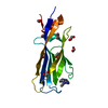
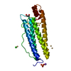
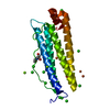
- PDB-1pv3: NMR Solution Structure of the Avian FAT-domain of Focal Adhesion ... -
+ Open data

- Basic information
| Entry | Database: PDB / ID: 1pv3 | ||||||
|---|---|---|---|---|---|---|---|
| Title | NMR Solution Structure of the Avian FAT-domain of Focal Adhesion Kinase | ||||||
 Components Components | Focal adhesion kinase 1 | ||||||
 Keywords Keywords | TRANSFERASE / Focal Adhesion Kinase / Helix Bundle / FAT-Domain | ||||||
| Function / homology |  Function and homology information Function and homology informationApoptotic cleavage of cellular proteins / NCAM signaling for neurite out-growth / RAF/MAP kinase cascade / Turbulent (oscillatory, disturbed) flow shear stress activates signaling by PIEZO1 and integrins in endothelial cells / RHO GTPases Activate WASPs and WAVEs / Regulation of actin dynamics for phagocytic cup formation / negative regulation of protein autophosphorylation / positive regulation of protein tyrosine kinase activity / radial glia-guided pyramidal neuron migration / calcium-dependent cysteine-type endopeptidase activity ...Apoptotic cleavage of cellular proteins / NCAM signaling for neurite out-growth / RAF/MAP kinase cascade / Turbulent (oscillatory, disturbed) flow shear stress activates signaling by PIEZO1 and integrins in endothelial cells / RHO GTPases Activate WASPs and WAVEs / Regulation of actin dynamics for phagocytic cup formation / negative regulation of protein autophosphorylation / positive regulation of protein tyrosine kinase activity / radial glia-guided pyramidal neuron migration / calcium-dependent cysteine-type endopeptidase activity / positive regulation of substrate-dependent cell migration, cell attachment to substrate / Integrin signaling / GRB2:SOS provides linkage to MAPK signaling for Integrins / : / MET activates PTK2 signaling / Extra-nuclear estrogen signaling / EPHB-mediated forward signaling / p130Cas linkage to MAPK signaling for integrins / VEGFA-VEGFR2 Pathway / signal complex assembly / response to pH / angiogenesis involved in wound healing / wound healing, spreading of cells / negative regulation of anoikis / positive regulation of protein binding / negative regulation of cell-substrate adhesion / positive regulation of focal adhesion assembly / regulation of cell adhesion / response to muscle stretch / molecular function activator activity / actin filament organization / non-membrane spanning protein tyrosine kinase activity / non-specific protein-tyrosine kinase / sarcolemma / integrin binding / epidermal growth factor receptor signaling pathway / protein autophosphorylation / protease binding / protein tyrosine kinase activity / cell cortex / positive regulation of cell migration / ciliary basal body / focal adhesion / positive regulation of cell population proliferation / centrosome / perinuclear region of cytoplasm / ATP binding / identical protein binding / nucleus / plasma membrane / cytoplasm Similarity search - Function | ||||||
| Biological species |  | ||||||
| Method | SOLUTION NMR / distance geometry simulated annealing | ||||||
 Authors Authors | Prutzman, K.C. / Gao, G. / King, M.L. / Iyer, V.V. / Mueller, G.A. / Schaller, M.D. / Campbell, S.L. | ||||||
 Citation Citation | Journal: STRUCTURE / Year: 2004 Title: The Focal Adhesion Targeting Domain of Focal Adhesion Kinase Contains a Hinge Region that Modulates Tyrosine 926 Phosphorylation. Authors: Prutzman, K.C. / Gao, G. / King, M.L. / Iyer, V.V. / Mueller, G.A. / Schaller, M.D. / Campbell, S.L. | ||||||
| History |
|
- Structure visualization
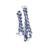
| Structure viewer | Molecule: MolmilJmol/JSmol |
|---|
- Downloads & links
-Download
| PDBx/mmCIF format | 1pv3.cif.gz | 877.2 KB | Display | PDBx/mmCIF format |
|---|---|---|---|---|
| PDB format | pdb1pv3.ent.gz | 735.7 KB | Display | PDB format |
| PDBx/mmJSON format | 1pv3.json.gz | Tree view | PDBx/mmJSON format | |
| Others |  Other downloads Other downloads |
-Validation report
| Arichive directory | https://data.pdbj.org/pub/pdb/validation_reports/pv/1pv3ftp://data.pdbj.org/pub/pdb/validation_reports/pv/1pv3 | HTTPS FTP |
|---|
-Related structure data
| Similar structure data |
|---|


-Links
 PDBj
PDBj
- Assembly
| Deposited unit | 
| |||||||||
|---|---|---|---|---|---|---|---|---|---|---|
| 1 |
| |||||||||
| NMR ensembles |
|
-Components
| #1: Protein | Mass: 15897.431 Da / Num. of mol.: 1 / Fragment: Focal Adhesion Targeting (FAT) Domain Source method: isolated from a genetically manipulated source Source: (gene. exp.)  |
|---|
-Experimental details
-Experiment
| Experiment | Method: SOLUTION NMR | ||||||||||||
|---|---|---|---|---|---|---|---|---|---|---|---|---|---|
| NMR experiment |
| ||||||||||||
| NMR details | Text: PDB entry 1K40 was used as a starting template for structure calculations. The structure was determined using triple-resonance NMR spectroscopy. |
- Sample preparation
| Details | Contents: 0.85 mM Focal Adhesion Targeting Domain U-15N,13C, 25 mM Tris-Maleate, 0.1 % NaN3, 1.0 uM PPACK, 0.5 mg/mL Pefabloc 90% H2O, 10% D2O Solvent system: 90% H2O/10% D2O |
|---|---|
| Sample conditions | Ionic strength: 150 mM NaCl / pH: 6 / Pressure: ambient / Temperature: 310 K |
-NMR measurement
| NMR spectrometer |
|
|---|
- Processing
| NMR software |
| ||||||||||||||||||||||||
|---|---|---|---|---|---|---|---|---|---|---|---|---|---|---|---|---|---|---|---|---|---|---|---|---|---|
| Refinement | Method: distance geometry simulated annealing / Software ordinal: 1 Details: 3049 total restraints: 1627 unambiguous NOE-derived distance constraints, 1078 ambiguous NOE-derived distance constraints, 83 dihedral angle restraints, 97 distance restraints from hydrogen ...Details: 3049 total restraints: 1627 unambiguous NOE-derived distance constraints, 1078 ambiguous NOE-derived distance constraints, 83 dihedral angle restraints, 97 distance restraints from hydrogen bonds, 164 residual dipolar coupling restraints | ||||||||||||||||||||||||
| NMR representative | Selection criteria: lowest energy | ||||||||||||||||||||||||
| NMR ensemble | Conformer selection criteria: structures with acceptable covalent geometry,structures with the lowest energy Conformers calculated total number: 25 / Conformers submitted total number: 20 |
 NMRPipe