 Movie
Movie Controller


+ Open data

- Basic information
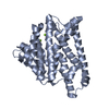
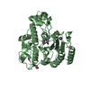
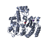
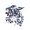
| Entry | Database: PDB / ID: 1ps1 | ||||||
|---|---|---|---|---|---|---|---|
| Title | PENTALENENE SYNTHASE | ||||||
 Components Components | PENTALENENE SYNTHASE | ||||||
 Keywords Keywords | ANTIBIOTIC BIOSYNTHESIS / SESQUITERPENE CYCLASE / LYASE | ||||||
| Function / homology |  Function and homology information Function and homology informationpentalenene synthase / pentalenene synthase activity / antibiotic biosynthetic process / metal ion binding Similarity search - Function | ||||||
| Biological species |  Streptomyces sp. (bacteria) Streptomyces sp. (bacteria) | ||||||
| Method |  X-RAY DIFFRACTION / SYNCHROTRON / MIR / Resolution: 2.6 Å X-RAY DIFFRACTION / SYNCHROTRON / MIR / Resolution: 2.6 Å | ||||||
 Authors Authors | Lesburg, C.A. / Christianson, D.W. | ||||||
 Citation Citation | Journal: Science / Year: 1997 Title: Crystal structure of pentalenene synthase: mechanistic insights on terpenoid cyclization reactions in biology. Authors: Lesburg, C.A. / Zhai, G. / Cane, D.E. / Christianson, D.W. #1: Journal: Protein Sci. / Year: 1995Title: Crystallization and Preliminary X-Ray Diffraction Analysis of Recombinant Pentalenene Synthase Authors: Lesburg, C.A. / Lloyd, M.D. / Cane, D.E. / Christianson, D.W. #2: Journal: Biochemistry / Year: 1994Title: Pentalenene Synthase. Purification, Molecular Cloning, Sequencing, and High-Level Expression in Escherichia Coli of a Terpenoid Cyclase from Streptomyces Uc5319 Authors: Cane, D.E. / Sohng, J.K. / Lamberson, C.R. / Rudnicki, S.M. / Wu, Z. / Lloyd, M.D. / Oliver, J.S. / Hubbard, B.R. | ||||||
| History |
|
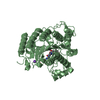
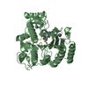
- Structure visualization
| Structure viewer | Molecule: MolmilJmol/JSmol |
|---|
- Downloads & links
-Download
| PDBx/mmCIF format | 1ps1.cif.gz | 130.3 KB | Display | PDBx/mmCIF format |
|---|---|---|---|---|
| PDB format | pdb1ps1.ent.gz | 103.7 KB | Display | PDB format |
| PDBx/mmJSON format | 1ps1.json.gz | Tree view | PDBx/mmJSON format | |
| Others |  Other downloads Other downloads |
-Validation report
| Arichive directory | https://data.pdbj.org/pub/pdb/validation_reports/ps/1ps1ftp://data.pdbj.org/pub/pdb/validation_reports/ps/1ps1 | HTTPS FTP |
|---|
-Related structure data
| Similar structure data |
|---|


-Links
 PDBj
PDBj
- Assembly
| Deposited unit | 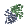
| ||||||||
|---|---|---|---|---|---|---|---|---|---|
| 1 | 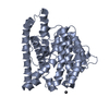
| ||||||||
| 2 | 
| ||||||||
| Unit cell |
| ||||||||
| Noncrystallographic symmetry (NCS) | NCS oper: (Code: given Matrix: (-0.298373, -0.228446, -0.926707), Vector: |
-Components
| #1: Protein | Mass: 38050.414 Da / Num. of mol.: 2 Source method: isolated from a genetically manipulated source Source: (gene. exp.) Streptomyces sp. (bacteria) / Strain: UC5319 / Production host: #2: Chemical | ChemComp-PBM / |   Mass: 252.304 Da / Num. of mol.: 1 / Source method: obtained synthetically / Formula: C3H9Pb Mass: 252.304 Da / Num. of mol.: 1 / Source method: obtained synthetically / Formula: C3H9Pb#3: Water | ChemComp-HOH / |  Mass: 18.015 Da / Num. of mol.: 66 / Source method: isolated from a natural source / Formula: H2O Mass: 18.015 Da / Num. of mol.: 66 / Source method: isolated from a natural source / Formula: H2OHas protein modification | Y | |
|---|
-Experimental details
-Experiment
| Experiment | Method: X-RAY DIFFRACTION / Number of used crystals: 1 |
|---|
- Sample preparation
| Crystal | Density Matthews: 3.4 Å3/Da / Density % sol: 64 % | |||||||||||||||||||||||||
|---|---|---|---|---|---|---|---|---|---|---|---|---|---|---|---|---|---|---|---|---|---|---|---|---|---|---|
| Crystal grow | pH: 7 Details: CRYSTALLIZED FROM 1.5M AMMONIUM SULFATE, 100 MM HEPES, PH 7.0 | |||||||||||||||||||||||||
| Crystal | *PLUS | |||||||||||||||||||||||||
| Crystal grow | *PLUS Method: vapor diffusion, hanging drop / Details: Lesburg, C.A., (1995) Protein Sci., 4, 2436. | |||||||||||||||||||||||||
| Components of the solutions | *PLUS
|
-Data collection
| Diffraction | Mean temperature: 100 K |
|---|---|
| Diffraction source | Source: SYNCHROTRON / Site: CHESS  / Beamline: A1 / Wavelength: 0.914 / Beamline: A1 / Wavelength: 0.914 |
| Detector | Type: PRINCETON 2K / Detector: CCD / Date: Sep 1, 1996 / Details: MIRRORS |
| Radiation | Monochromator: SI(111) / Monochromatic (M) / Laue (L): M / Scattering type: x-ray |
| Radiation wavelength | Wavelength: 0.914 Å / Relative weight: 1 |
| Reflection | Resolution: 2.6→35 Å / Num. obs: 28128 / % possible obs: 87.5 % / Observed criterion σ(I): 0 / Redundancy: 2.5 % / Rsym value: 0.056 / Net I/σ(I): 13.8 |
| Reflection shell | Resolution: 2.6→2.69 Å / Rsym value: 0.158 / % possible all: 57.7 |
| Reflection | *PLUS Num. measured all: 70683 / Rmerge(I) obs: 0.056 |
| Reflection shell | *PLUS Rmerge(I) obs: 0.158 |
- Processing
| Software |
| ||||||||||||||||||||||||||||||||||||||||||||||||||||||||||||||||||||||||||||||||
|---|---|---|---|---|---|---|---|---|---|---|---|---|---|---|---|---|---|---|---|---|---|---|---|---|---|---|---|---|---|---|---|---|---|---|---|---|---|---|---|---|---|---|---|---|---|---|---|---|---|---|---|---|---|---|---|---|---|---|---|---|---|---|---|---|---|---|---|---|---|---|---|---|---|---|---|---|---|---|---|---|---|
| Refinement | Method to determine structure: MIR / Resolution: 2.6→20 Å / Rfactor Rfree error: 0.007 / Data cutoff high absF: 100000 / Data cutoff low absF: 0.1 / Isotropic thermal model: RESTRAINED / Cross valid method: THROUGHOUT / σ(F): 3
| ||||||||||||||||||||||||||||||||||||||||||||||||||||||||||||||||||||||||||||||||
| Displacement parameters | Biso mean: 52.1 Å2 | ||||||||||||||||||||||||||||||||||||||||||||||||||||||||||||||||||||||||||||||||
| Refine analyze |
| ||||||||||||||||||||||||||||||||||||||||||||||||||||||||||||||||||||||||||||||||
| Refinement step | Cycle: LAST / Resolution: 2.6→20 Å
| ||||||||||||||||||||||||||||||||||||||||||||||||||||||||||||||||||||||||||||||||
| Refine LS restraints |
| ||||||||||||||||||||||||||||||||||||||||||||||||||||||||||||||||||||||||||||||||
| Refine LS restraints NCS | NCS model details: RESTRAINTS / Rms dev Biso : 17.4 Å2 / Rms dev position: 0.43 Å / Weight Biso : 44.4 / Weight position: 9 | ||||||||||||||||||||||||||||||||||||||||||||||||||||||||||||||||||||||||||||||||
| LS refinement shell | Resolution: 2.6→2.66 Å / Rfactor Rfree error: 0.058 / Total num. of bins used: 15
| ||||||||||||||||||||||||||||||||||||||||||||||||||||||||||||||||||||||||||||||||
| Xplor file |
| ||||||||||||||||||||||||||||||||||||||||||||||||||||||||||||||||||||||||||||||||
| Software | *PLUS Name: X-PLOR / Version: 3.1 / Classification: refinement | ||||||||||||||||||||||||||||||||||||||||||||||||||||||||||||||||||||||||||||||||
| Refine LS restraints | *PLUS
|
