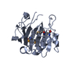
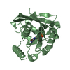
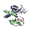
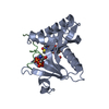
- PDB-1pjz: Solution structure of thiopurine methyltransferase from Pseudomon... -
+
データを開く
IDまたはキーワード:
読み込み中...
-
基本情報
登録情報
データベース: PDB / ID: 1pjz
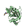
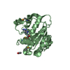
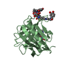
タイトル
Solution structure of thiopurine methyltransferase from Pseudomonas syringae
要素
Thiopurine S-methyltransferase
キーワード
TRANSFERASE / methyltransferase / polymorphism / S-adenosylmethionine / drug metabolism
機能・相同性
機能・相同性情報
thiopurine S-methyltransferase / thiopurine S-methyltransferase activity / response to tellurium ion / response to metal ion / methylation / cytoplasm 類似検索 - 分子機能
Thiopurine S-methyltransferase, Se/Te detoxification / Thiopurine S-methyltransferase / Thiopurine S-methyltransferase (TPMT) / TPMT family / Thiopurine or thiol or thiocyanate S-methyltransferase (TPMT) family profile. / Vaccinia Virus protein VP39 / S-adenosyl-L-methionine-dependent methyltransferase superfamily / Rossmann fold / 3-Layer(aba) Sandwich / Alpha Beta 類似検索 - ドメイン・相同性
内容: 20 mM Potassium Phosphate, 150 mM NaCl, 0.05% NaN3, 1 uM lupeptin, 1 uM pepstatin, 1 uM PMSF, 1.5 mM protein, uniform (random) labeling with 13C, 15N at known labeling 溶媒系: 95% H2O/5% D2O
試料状態
イオン強度: 150 mM NaCL / pH: 6.8 / 圧: 1 atm / 温度: 293 K
 ムービー
ムービー コントローラー


 データを開く
 基本情報
基本情報 要素
要素 キーワード
キーワード 機能・相同性情報
 Pseudomonas syringae pv. pisi (バクテリア)
 データ登録者
 引用
引用 構造の表示
構造の表示 ダウンロードとリンク
 その他のダウンロード
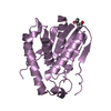


 PDBj
PDBj 集合体
集合体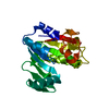
 試料調製
試料調製 解析
解析 NMRPipe