 Movie
Movie Controller


+ Open data

- Basic information
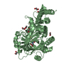
| Entry | Database: PDB / ID: 1pin | |||||||||
|---|---|---|---|---|---|---|---|---|---|---|
| Title | PIN1 PEPTIDYL-PROLYL CIS-TRANS ISOMERASE FROM HOMO SAPIENS | |||||||||
 Components Components | PEPTIDYL-PROLYL CIS-TRANS ISOMERASE | |||||||||
 Keywords Keywords | ISOMERASE / PEPTIDYL-PROLYL CIS-TRANS ISOMERASE / ROTAMASE / COMPLEX (ISOMERASE-DIPEPTIDE) | |||||||||
| Function / homology |  Function and homology information Function and homology informationcis-trans isomerase activity / phosphothreonine residue binding / negative regulation of brown fat cell differentiation / negative regulation of cell motility / ubiquitin ligase activator activity / regulation of protein localization to nucleus / GTPase activating protein binding / mitogen-activated protein kinase kinase binding / protein peptidyl-prolyl isomerization / : ...cis-trans isomerase activity / phosphothreonine residue binding / negative regulation of brown fat cell differentiation / negative regulation of cell motility / ubiquitin ligase activator activity / regulation of protein localization to nucleus / GTPase activating protein binding / mitogen-activated protein kinase kinase binding / protein peptidyl-prolyl isomerization / : / regulation of mitotic nuclear division / negative regulation of SMAD protein signal transduction / PI5P Regulates TP53 Acetylation / negative regulation of amyloid-beta formation / cytoskeletal motor activity / RHO GTPases Activate NADPH Oxidases / phosphoserine residue binding / postsynaptic cytosol / Rho protein signal transduction / regulation of cytokinesis / peptidylprolyl isomerase / peptidyl-prolyl cis-trans isomerase activity / Negative regulators of DDX58/IFIH1 signaling / negative regulation of transforming growth factor beta receptor signaling pathway / phosphoprotein binding / regulation of protein stability / synapse organization / beta-catenin binding / negative regulation of protein catabolic process / negative regulation of ERK1 and ERK2 cascade / ISG15 antiviral mechanism / protein destabilization / tau protein binding / positive regulation of protein phosphorylation / neuron differentiation / positive regulation of canonical Wnt signaling pathway / regulation of gene expression / midbody / cellular response to hypoxia / Regulation of TP53 Activity through Phosphorylation / response to hypoxia / protein stabilization / nuclear speck / ciliary basal body / glutamatergic synapse / positive regulation of transcription by RNA polymerase II / nucleoplasm / nucleus / cytoplasm / cytosol Similarity search - Function | |||||||||
| Biological species |  Homo sapiens (human) Homo sapiens (human) | |||||||||
| Method |  X-RAY DIFFRACTION / SYNCHROTRON / RIGID BODY REFINEMENT USING MIRAS DERIVED STRUCTURE / Resolution: 1.35 Å X-RAY DIFFRACTION / SYNCHROTRON / RIGID BODY REFINEMENT USING MIRAS DERIVED STRUCTURE / Resolution: 1.35 Å | |||||||||
 Authors Authors | Noel, J.P. / Ranganathan, R. / Hunter, T. | |||||||||
 Citation Citation | Journal: Cell(Cambridge,Mass.) / Year: 1997 Title: Structural and functional analysis of the mitotic rotamase Pin1 suggests substrate recognition is phosphorylation dependent. Authors: Ranganathan, R. / Lu, K.P. / Hunter, T. / Noel, J.P. #1: Journal: Nature / Year: 1996Title: A Human Peptidyl-Prolyl Isomerase Essential for Regulation of Mitosis Authors: Lu, K.P. / Hanes, S.D. / Hunter, T. | |||||||||
| History |
|
- Structure visualization
| Structure viewer | Molecule: MolmilJmol/JSmol |
|---|
- Downloads & links
-Download
| PDBx/mmCIF format | 1pin.cif.gz | 50.8 KB | Display | PDBx/mmCIF format |
|---|---|---|---|---|
| PDB format | pdb1pin.ent.gz | 35.6 KB | Display | PDB format |
| PDBx/mmJSON format | 1pin.json.gz | Tree view | PDBx/mmJSON format | |
| Others |  Other downloads Other downloads |
-Validation report
| Arichive directory | https://data.pdbj.org/pub/pdb/validation_reports/pi/1pinftp://data.pdbj.org/pub/pdb/validation_reports/pi/1pin | HTTPS FTP |
|---|
-Related structure data
| Similar structure data |
|---|


-Links
 PDBj
PDBj


- Assembly
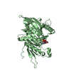
| Deposited unit | 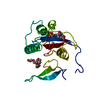
| ||||||||
|---|---|---|---|---|---|---|---|---|---|
| 1 | 
| ||||||||
| Unit cell |
|
-Components
-Protein , 1 types, 1 molecules A
| #1: Protein | Mass: 18271.309 Da / Num. of mol.: 1 Source method: isolated from a genetically manipulated source Source: (gene. exp.) Homo sapiens (human)Description: USING THE HUMAN GENE, THE PROTEIN WAS OVEREXPRESSED IN ESCHERICHIA COLI. THE GENE FOR HUMAN PIN1 WAS INSERTED INTO THE NCOI/BAMHI SITES OF PLASMID PET28A(+) - NOVAGEN -, TRANSFORMED INTO ...Description: USING THE HUMAN GENE, THE PROTEIN WAS OVEREXPRESSED IN ESCHERICHIA COLI. THE GENE FOR HUMAN PIN1 WAS INSERTED INTO THE NCOI/BAMHI SITES OF PLASMID PET28A(+) - NOVAGEN -, TRANSFORMED INTO E. COLI BL21(DE3), AND EXPRESSED AT 20 DEGREES CELSIUS AS A 6HIS N-TERMINAL FUSION PROTEIN. FOLLOWING PURIFICATION USING A NI-NTA RESIN, THE 6HIS FUSION WAS REMOVED BY THROMBIN DIGESTION. Cell line: HELA CELL / Gene: PIN1 / Plasmid: PET28A(+) / Species (production host): Escherichia coli / Cellular location (production host): CYTOPLASM / Production host:  |
|---|
-Non-polymers , 5 types, 209 molecules 


| #2: Chemical | ChemComp-ALA /  Type: L-peptide linking / Mass: 89.093 Da / Num. of mol.: 1 / Source method: obtained synthetically / Formula: C3H7NO2 Type: L-peptide linking / Mass: 89.093 Da / Num. of mol.: 1 / Source method: obtained synthetically / Formula: C3H7NO2 | ||
|---|---|---|---|
| #3: Chemical | ChemComp-PRO /  Type: L-peptide linking / Mass: 115.130 Da / Num. of mol.: 1 / Source method: obtained synthetically / Formula: C5H9NO2 Type: L-peptide linking / Mass: 115.130 Da / Num. of mol.: 1 / Source method: obtained synthetically / Formula: C5H9NO2 | ||
| #4: Chemical | ChemComp-SO4 /  Mass: 96.063 Da / Num. of mol.: 1 / Source method: obtained synthetically / Formula: SO4 Mass: 96.063 Da / Num. of mol.: 1 / Source method: obtained synthetically / Formula: SO4 | ||
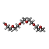
| #5: Chemical |  Mass: 252.305 Da / Num. of mol.: 2 / Source method: obtained synthetically / Formula: C11H24O6 Mass: 252.305 Da / Num. of mol.: 2 / Source method: obtained synthetically / Formula: C11H24O6#6: Water | ChemComp-HOH / | Mass: 18.015 Da / Num. of mol.: 204 / Source method: isolated from a natural source / Formula: H2O |
-Details
| Nonpolymer details | RESIDUES 201 AND 202 WITH FORMS A DIPEPTIDE (ALA-PRO) THAT IS BOUND TO THE PEPTIDYL-PROLYL CIS-TRANS ISOMERASE |
|---|
-Experimental details
-Experiment
| Experiment | Method: X-RAY DIFFRACTION / Number of used crystals: 1 |
|---|
- Sample preparation
| Crystal | Density Matthews: 2.24 Å3/Da / Density % sol: 42 % | ||||||||||||||||||||||||||||||||||||
|---|---|---|---|---|---|---|---|---|---|---|---|---|---|---|---|---|---|---|---|---|---|---|---|---|---|---|---|---|---|---|---|---|---|---|---|---|---|
| Crystal grow | Temperature: 277 K / pH: 7.5 Details: PROTEIN WAS CRYSTALLIZED AT 4 DEGREES CELSIUS FROM 2.4 M (NH4)2SO4, 1% (V/V) PEG 400, 0.1 M NA-HEPES, PH 7.5. PRIOR TO DATA COLLECTION, THE CRYSTALS WERE TRANSFERRED TO SOLUTIONS OF 40 % ...Details: PROTEIN WAS CRYSTALLIZED AT 4 DEGREES CELSIUS FROM 2.4 M (NH4)2SO4, 1% (V/V) PEG 400, 0.1 M NA-HEPES, PH 7.5. PRIOR TO DATA COLLECTION, THE CRYSTALS WERE TRANSFERRED TO SOLUTIONS OF 40 % (V/V) PEG 400, 0.1 M NA-HEPES, PH 7.5 CONTAINING 0.05 M ALANINE-PROLINE DIPEPTIDE., temperature 277K | ||||||||||||||||||||||||||||||||||||
| Crystal | *PLUS | ||||||||||||||||||||||||||||||||||||
| Crystal grow | *PLUS Temperature: 4 ℃ / Method: vapor diffusion, hanging drop | ||||||||||||||||||||||||||||||||||||
| Components of the solutions | *PLUS
|
-Data collection
| Diffraction | Mean temperature: 100 K |
|---|---|
| Diffraction source | Source: SYNCHROTRON / Site: SSRL  / Beamline: BL7-1 / Wavelength: 1.08 / Beamline: BL7-1 / Wavelength: 1.08 |
| Detector | Type: MARRESEARCH / Detector: IMAGE PLATE / Date: Mar 1, 1996 |
| Radiation | Monochromatic (M) / Laue (L): M / Scattering type: x-ray |
| Radiation wavelength | Wavelength: 1.08 Å / Relative weight: 1 |
| Reflection | Resolution: 1.35→25 Å / Num. obs: 33672 / % possible obs: 95.5 % / Redundancy: 4.5 % / Rsym value: 0.053 / Net I/σ(I): 18 |
| Reflection shell | Resolution: 1.35→1.39 Å / Mean I/σ(I) obs: 2 / Rsym value: 0.592 / % possible all: 69 |
| Reflection | *PLUS Rmerge(I) obs: 0.053 |
| Reflection shell | *PLUS % possible obs: 69 % / Rmerge(I) obs: 0.592 |
- Processing
| Software |
| ||||||||||||||||||||||||||||||||||||||||||||||||||||||||||||||||||||||||||||||||
|---|---|---|---|---|---|---|---|---|---|---|---|---|---|---|---|---|---|---|---|---|---|---|---|---|---|---|---|---|---|---|---|---|---|---|---|---|---|---|---|---|---|---|---|---|---|---|---|---|---|---|---|---|---|---|---|---|---|---|---|---|---|---|---|---|---|---|---|---|---|---|---|---|---|---|---|---|---|---|---|---|---|
| Refinement | Method to determine structure: RIGID BODY REFINEMENT USING MIRAS DERIVED STRUCTURE Resolution: 1.35→6 Å / Rfactor Rfree error: 0.01 / Data cutoff high absF: 1000000 / Data cutoff low absF: 0.1 / Isotropic thermal model: RESTRAINED / Cross valid method: THROUGHOUT / σ(F): 0 Details: RESIDUES 1 - 5 AND 40 - 44 (WHICH LINK THE WW DOMAIN TO THE PPIASE DOMAIN) WERE NOT VISIBLE IN THE FINAL ELECTRON DENSITY MAP AND SO WERE NOT MODELLED.
| ||||||||||||||||||||||||||||||||||||||||||||||||||||||||||||||||||||||||||||||||
| Displacement parameters | Biso mean: 20 Å2 | ||||||||||||||||||||||||||||||||||||||||||||||||||||||||||||||||||||||||||||||||
| Refinement step | Cycle: LAST / Resolution: 1.35→6 Å
| ||||||||||||||||||||||||||||||||||||||||||||||||||||||||||||||||||||||||||||||||
| Refine LS restraints |
| ||||||||||||||||||||||||||||||||||||||||||||||||||||||||||||||||||||||||||||||||
| LS refinement shell | Resolution: 1.35→1.39 Å / Rfactor Rfree error: 0.05 / Total num. of bins used: 12
| ||||||||||||||||||||||||||||||||||||||||||||||||||||||||||||||||||||||||||||||||
| Xplor file |
| ||||||||||||||||||||||||||||||||||||||||||||||||||||||||||||||||||||||||||||||||
| Software | *PLUS Name: X-PLOR / Version: 3.851 / Classification: refinement | ||||||||||||||||||||||||||||||||||||||||||||||||||||||||||||||||||||||||||||||||
| Refine LS restraints | *PLUS
|
