 Movie
Movie Controller

[English] 日本語
 Yorodumi
- PDB-1pek: STRUCTURE OF THE COMPLEX OF PROTEINASE K WITH A SUBSTRATE-ANALOGU... -
+ Open data

- Basic information
| Entry | Database: PDB / ID: 1pek | ||||||
|---|---|---|---|---|---|---|---|
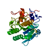
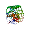
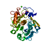
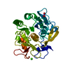
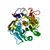
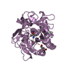
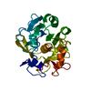
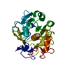
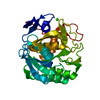
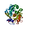
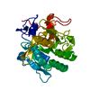
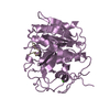
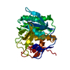
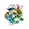
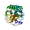
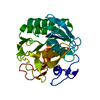
| Title | STRUCTURE OF THE COMPLEX OF PROTEINASE K WITH A SUBSTRATE-ANALOGUE HEXA-PEPTIDE INHIBITOR AT 2.2 ANGSTROMS RESOLUTION | ||||||
 Components Components |
| ||||||
 Keywords Keywords | HYDROLASE/HYDROLASE INHIBITOR / HYDROLASE-HYDROLASE INHIBITOR complex | ||||||
| Function / homology |  Function and homology information Function and homology informationpeptidase K / serine-type endopeptidase activity / proteolysis / extracellular region / metal ion binding Similarity search - Function | ||||||
| Biological species |  Tritirachium album (fungus) Tritirachium album (fungus) | ||||||
| Method |  X-RAY DIFFRACTION / Resolution: 2.2 Å X-RAY DIFFRACTION / Resolution: 2.2 Å | ||||||
 Authors Authors | Betzel, C. / Singh, T.P. / Visanji, M. / Peters, K. / Fittkau, S. / Saenger, W. / Wilson, K.S. | ||||||
 Citation Citation | Journal: J.Biol.Chem. / Year: 1993 Title: Structure of the complex of proteinase K with a substrate analogue hexapeptide inhibitor at 2.2-A resolution. Authors: Betzel, C. / Singh, T.P. / Visanji, M. / Peters, K. / Fittkau, S. / Saenger, W. / Wilson, K.S. #1: Journal: Eur.J.Biochem. / Year: 1988Title: Three-Dimensional Structure of Proteinase K at 0.15-Nm Resolution Authors: Betzel, C. / Pal, G.P. / Saenger, W. | ||||||
| History |
|
- Structure visualization
| Structure viewer | Molecule: MolmilJmol/JSmol |
|---|
- Downloads & links
-Download
| PDBx/mmCIF format | 1pek.cif.gz | 71.8 KB | Display | PDBx/mmCIF format |
|---|---|---|---|---|
| PDB format | pdb1pek.ent.gz | 48.9 KB | Display | PDB format |
| PDBx/mmJSON format | 1pek.json.gz | Tree view | PDBx/mmJSON format | |
| Others |  Other downloads Other downloads |
-Validation report
| Arichive directory | https://data.pdbj.org/pub/pdb/validation_reports/pe/1pekftp://data.pdbj.org/pub/pdb/validation_reports/pe/1pek | HTTPS FTP |
|---|
-Related structure data
| Similar structure data |
|---|


-Links
 PDBj
PDBj

- Assembly
| Deposited unit | 
| ||||||||
|---|---|---|---|---|---|---|---|---|---|
| 1 |
| ||||||||
| Unit cell |
| ||||||||
| Atom site foot note | 1: ASN E 168 - TYR E 169 OMEGA = 359.43 PEPTIDE BOND DEVIATES SIGNIFICANTLY FROM TRANS CONFORMATION 2: SER E 170 - PRO E 171 OMEGA = 75.63 PEPTIDE BOND DEVIATES SIGNIFICANTLY FROM TRANS CONFORMATION 3: PRO C 3 - PHE C 4 OMEGA = 359.45 PEPTIDE BOND DEVIATES SIGNIFICANTLY FROM TRANS CONFORMATION 4: RESIDUE ALA D 5 IS D-ALANINE. |
-Components
| #1: Protein | Mass: 28958.838 Da / Num. of mol.: 1 / Source method: isolated from a natural source / Source: (natural) Tritirachium album (fungus) / References: UniProt: P06873, peptidase K | ||||
|---|---|---|---|---|---|
| #2: Protein/peptide | Mass: 430.496 Da / Num. of mol.: 1 / Source method: obtained synthetically Details: THE COMPLETE PEPTIDE IS N-AC-PRO-ALA-PRO-PHE-D-ALA-ALA-NH2 AND IS HYDROLYSED AND REPRESENTED AS CHAINS C AND D | ||||
| #3: Protein/peptide | Mass: 158.179 Da / Num. of mol.: 1 / Source method: obtained synthetically Details: THE COMPLETE PEPTIDE IS N-AC-PRO-ALA-PRO-PHE-D-ALA-ALA-NH2 AND IS HYDROLYSED AND REPRESENTED AS CHAINS C AND D | ||||
| #4: Water | ChemComp-HOH /  Mass: 18.015 Da / Num. of mol.: 188 / Source method: isolated from a natural source / Formula: H2O Mass: 18.015 Da / Num. of mol.: 188 / Source method: isolated from a natural source / Formula: H2O | ||||
| Compound details | THE HEXAPEPTID| Has protein modification | Y | Sequence details | SEQUENCE ADVISORY NOTICE: DIFFERENCE BETWEEN SWISS-PROT AND PDB SEQUENCE. SWISS-PROT ENTRY NAME: ...SEQUENCE ADVISORY NOTICE: DIFFERENCE | |
-Experimental details
-Experiment
| Experiment | Method: X-RAY DIFFRACTION |
|---|
- Sample preparation
| Crystal | Density Matthews: 2.13 Å3/Da / Density % sol: 42.16 % | ||||||||||||||||||||||||
|---|---|---|---|---|---|---|---|---|---|---|---|---|---|---|---|---|---|---|---|---|---|---|---|---|---|
| Crystal grow | *PLUS Temperature: 16 ℃ / pH: 6.5 / Method: unknown | ||||||||||||||||||||||||
| Components of the solutions | *PLUS
|
-Data collection
| Radiation | Scattering type: x-ray |
|---|---|
| Radiation wavelength | Relative weight: 1 |
| Reflection | *PLUS Highest resolution: 2.2 Å / Num. obs: 12725 / % possible obs: 95 % / Num. measured all: 44167 / Rmerge(I) obs: 0.08 |
- Processing
| Software | Name: PROLSQ / Classification: refinement | ||||||||||||||||||||||||||||||||||||||||||||||||||||||||||||||||||||||||||||||||||||
|---|---|---|---|---|---|---|---|---|---|---|---|---|---|---|---|---|---|---|---|---|---|---|---|---|---|---|---|---|---|---|---|---|---|---|---|---|---|---|---|---|---|---|---|---|---|---|---|---|---|---|---|---|---|---|---|---|---|---|---|---|---|---|---|---|---|---|---|---|---|---|---|---|---|---|---|---|---|---|---|---|---|---|---|---|---|
| Refinement | Resolution: 2.2→8 Å / σ(F): 0 /
| ||||||||||||||||||||||||||||||||||||||||||||||||||||||||||||||||||||||||||||||||||||
| Refinement step | Cycle: LAST / Resolution: 2.2→8 Å
| ||||||||||||||||||||||||||||||||||||||||||||||||||||||||||||||||||||||||||||||||||||
| Refine LS restraints |
| ||||||||||||||||||||||||||||||||||||||||||||||||||||||||||||||||||||||||||||||||||||
| Refinement | *PLUS Highest resolution: 2.2 Å / Lowest resolution: 8 Å / Num. reflection all: 12725 / σ(F): 0 / Rfactor all: 0.165 | ||||||||||||||||||||||||||||||||||||||||||||||||||||||||||||||||||||||||||||||||||||
| Solvent computation | *PLUS | ||||||||||||||||||||||||||||||||||||||||||||||||||||||||||||||||||||||||||||||||||||
| Displacement parameters | *PLUS Biso mean: 11.9 Å2 |
