 Movie
Movie Controller

[English] 日本語
 Yorodumi
- PDB-1ouc: CONTRIBUTION OF HYDROPHOBIC RESIDUES TO THE STABILITY OF HUMAN LY... -
+ Open data

- Basic information
| Entry | Database: PDB / ID: 1ouc | ||||||
|---|---|---|---|---|---|---|---|
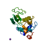
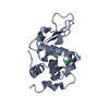
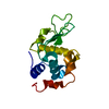
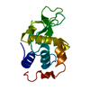
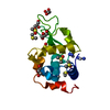
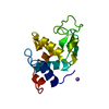
| Title | CONTRIBUTION OF HYDROPHOBIC RESIDUES TO THE STABILITY OF HUMAN LYSOZYME: X-RAY STRUCTURE OF THE V110A MUTANT | ||||||
 Components Components | LYSOZYME | ||||||
 Keywords Keywords | HYDROLASE (O-GLYCOSYL) / AMYLOID / DISEASE MUTATION | ||||||
| Function / homology |  Function and homology information Function and homology informationantimicrobial humoral response / Antimicrobial peptides / metabolic process / specific granule lumen / azurophil granule lumen / tertiary granule lumen / lysozyme / lysozyme activity / killing of cells of another organism / defense response to Gram-negative bacterium ...antimicrobial humoral response / Antimicrobial peptides / metabolic process / specific granule lumen / azurophil granule lumen / tertiary granule lumen / lysozyme / lysozyme activity / killing of cells of another organism / defense response to Gram-negative bacterium / defense response to Gram-positive bacterium / defense response to bacterium / inflammatory response / Amyloid fiber formation / Neutrophil degranulation / extracellular space / extracellular exosome / extracellular region / identical protein bindingSimilarity search - Function | ||||||
| Biological species |  Homo sapiens (human) Homo sapiens (human) | ||||||
| Method | X-RAY DIFFRACTION / Resolution: 1.8 Å | ||||||
 Authors Authors | Takano, K. / Yamagata, Y. / Fujii, S. / Yutani, K. | ||||||
 Citation Citation | Journal: Biochemistry / Year: 1997 Title: Contribution of the hydrophobic effect to the stability of human lysozyme: calorimetric studies and X-ray structural analyses of the nine valine to alanine mutants. Authors: Takano, K. / Yamagata, Y. / Fujii, S. / Yutani, K. #1: Journal: J.Mol.Biol. / Year: 1995Title: Contribution of Hydrophobic Residues to the Stability of Human Lysozyme: Calorimetric Studies and X-Ray Structural Analysis of the Five Isoleucine to Valine Mutants Authors: Takano, K. / Ogasahara, K. / Kaneda, H. / Yamagata, Y. / Fujii, S. / Kanaya, E. / Kikuchi, M. / Oobatake, M. / Yutani, K. | ||||||
| History |
|
- Structure visualization
| Structure viewer | Molecule: MolmilJmol/JSmol |
|---|
- Downloads & links
-Download
| PDBx/mmCIF format | 1ouc.cif.gz | 38.8 KB | Display | PDBx/mmCIF format |
|---|---|---|---|---|
| PDB format | pdb1ouc.ent.gz | 28.9 KB | Display | PDB format |
| PDBx/mmJSON format | 1ouc.json.gz | Tree view | PDBx/mmJSON format | |
| Others |  Other downloads Other downloads |
-Validation report
| Arichive directory | https://data.pdbj.org/pub/pdb/validation_reports/ou/1oucftp://data.pdbj.org/pub/pdb/validation_reports/ou/1ouc | HTTPS FTP |
|---|
-Related structure data
| Related structure data | 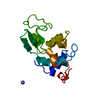 1oubC  1oudC  1oueC 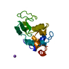 1oufC 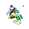 1ougC 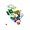 1ouhC  1ouiC  1oujC C: citing same article ( |
|---|---|
| Similar structure data |


-Links
 PDBj
PDBj


- Assembly
| Deposited unit | 
| ||||||||
|---|---|---|---|---|---|---|---|---|---|
| 1 |
| ||||||||
| Unit cell |
|
-Components
| #1: Protein | Mass: 14692.639 Da / Num. of mol.: 1 / Mutation: V110A Source method: isolated from a genetically manipulated source Source: (gene. exp.) Homo sapiens (human) / Gene: HUMAN LYSOZYME WITH VAL 110 / Plasmid: PERI8602Gene (production host): HUMAN LYSOZYME WITH VAL 110 REPLACED BY ALA Production host:  Saccharomyces cerevisiae (brewer's yeast) / Strain (production host): AH22R- / References: UniProt: P61626, lysozyme Saccharomyces cerevisiae (brewer's yeast) / Strain (production host): AH22R- / References: UniProt: P61626, lysozyme |
|---|---|
| #2: Chemical | ChemComp-NA /   Mass: 22.990 Da / Num. of mol.: 1 / Source method: obtained synthetically / Formula: Na Mass: 22.990 Da / Num. of mol.: 1 / Source method: obtained synthetically / Formula: Na |
| #3: Water | ChemComp-HOH / Water Mass: 18.015 Da / Num. of mol.: 206 / Source method: isolated from a natural source / Formula: H2O Mass: 18.015 Da / Num. of mol.: 206 / Source method: isolated from a natural source / Formula: H2O |
-Experimental details
-Experiment
| Experiment | Method: X-RAY DIFFRACTION / Number of used crystals: 1 |
|---|
- Sample preparation
| Crystal | Density Matthews: 1.99 Å3/Da / Density % sol: 38.23 % | ||||||||||||||||||||||||
|---|---|---|---|---|---|---|---|---|---|---|---|---|---|---|---|---|---|---|---|---|---|---|---|---|---|
| Crystal grow | pH: 4.5 / Details: pH 4.5 | ||||||||||||||||||||||||
| Crystal grow | *PLUS Temperature: 10 ℃ / Method: vapor diffusion, hanging drop / Details: Takano, K., (1995) J.Mol.Biol., 254, 62. | ||||||||||||||||||||||||
| Components of the solutions | *PLUS
|
-Data collection
| Diffraction | Mean temperature: 283 K |
|---|---|
| Diffraction source | Source: ROTATING ANODE / Type: RIGAKU RUH3R / Wavelength: 1.5418 |
| Detector | Type: RIGAKU RAXIS IIC / Detector: IMAGE PLATE / Date: Oct 4, 1994 |
| Radiation | Monochromatic (M) / Laue (L): M / Scattering type: x-ray |
| Radiation wavelength | Wavelength: 1.5418 Å / Relative weight: 1 |
| Reflection | Highest resolution: 1.8 Å / Num. obs: 10973 / % possible obs: 95.9 % / Observed criterion σ(I): 1 / Redundancy: 3.16 % / Rmerge(I) obs: 0.034 |
| Reflection | *PLUS Num. measured all: 34707 |
- Processing
| Software |
| ||||||||||||||||||||||||||||||||||||||||||||||||||||||||||||
|---|---|---|---|---|---|---|---|---|---|---|---|---|---|---|---|---|---|---|---|---|---|---|---|---|---|---|---|---|---|---|---|---|---|---|---|---|---|---|---|---|---|---|---|---|---|---|---|---|---|---|---|---|---|---|---|---|---|---|---|---|---|
| Refinement | Resolution: 1.8→8 Å / σ(F): 3
| ||||||||||||||||||||||||||||||||||||||||||||||||||||||||||||
| Refinement step | Cycle: LAST / Resolution: 1.8→8 Å
| ||||||||||||||||||||||||||||||||||||||||||||||||||||||||||||
| Refine LS restraints |
| ||||||||||||||||||||||||||||||||||||||||||||||||||||||||||||
| Software | *PLUS Name: X-PLOR / Classification: refinement | ||||||||||||||||||||||||||||||||||||||||||||||||||||||||||||
| Refine LS restraints | *PLUS
|
