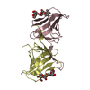
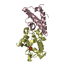
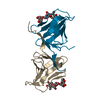
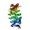
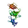
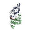
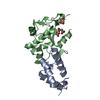
SOLUTION NMR / THE STRUCTURE OF THE COMPLEX WAS CALCULATED AS FOLLOWS. FIRST THE STRUCTURE OF THE DIMERIC LACHP62-V52C WAS CALCULATED USING ONLY PROTEIN NMR RESTRAINTS. THE 100 BEST STRUCTURES WERE SELECTED, DOCKED ONTO THE NONSPECIFIC LAC OPERATOR B-DNA USING SIMULATED ANNEALING. DISTANCE, PLANARITY RESTRAINTS FOR THE DNA WERE INCORPORATED IN ORDER TO KEEP DNA CLOSE TO B-DNA CONFORMATION.
Mass: 5514.603 Da / Num. of mol.: 2 / Source method: obtained synthetically
#2: Protein
Lactoseoperonrepressor
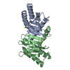
Mass: 6822.755 Da / Num. of mol.: 2 / Fragment: N-terminal DNA-binding domain, residues 1-62 / Mutation: V52C Source method: isolated from a genetically manipulated source Source: (gene. exp.) Escherichia coli (E. coli) / Gene: LACI / Plasmid: PET-HP62-V52C / Production host: Escherichia coli (E. coli) / Strain (production host): DH9 / References: UniProt: P03023
Has protein modification
Y
-
Experimental details
-
Experiment
Experiment
Method: SOLUTION NMR
NMR experiment
Conditions-ID
Experiment-ID
Solution-ID
Type
1
1
1
3D 15N-separated NOESY
1
2
1
3D 13C-separated NOESY
1
3
1
13C-15N double-half NOESY filter
NMR details
Text: THIS STRUCTURE WAS DETERMINED USING STANDARD 2D AND 3D HOMO- AND HETERONUCLEAR TECHNIQUES. 13C-15N LABELED PROTEIN AND UNLABELED NUCLEOTIDE WERE USED. IN ADDITION ISOTOPE FILTER EXPERIMENTS ...Text: THIS STRUCTURE WAS DETERMINED USING STANDARD 2D AND 3D HOMO- AND HETERONUCLEAR TECHNIQUES. 13C-15N LABELED PROTEIN AND UNLABELED NUCLEOTIDE WERE USED. IN ADDITION ISOTOPE FILTER EXPERIMENTS WERE APPLIED TO OBTAIN ADDITIONAL ASSIGNMENTS AND TO ASSIGN INTER-MOLECULAR NOES. FOR FURTHER DETAILS SEE THE REFERENCE DESCRIBING THE STRUCTURES.
Protocol: SINGLE WAVELENGTH / Monochromatic (M) / Laue (L): M
Radiation wavelength
Relative weight: 1
NMR spectrometer
Type: Bruker DRX / Manufacturer: Bruker / Model: DRX / Field strength: 750 MHz
-
Processing
NMR software
Name
Version
Developer
Classification
XwinNMR
3
bruker
collection
NMRPipe
2.1
delaglio
processing
NMRView
5.0.3
Johnson
dataanalysis
CNS
1.1
brunger
structuresolution
CNS
1.1
brunger
refinement
Refinement
Method: THE STRUCTURE OF THE COMPLEX WAS CALCULATED AS FOLLOWS. FIRST THE STRUCTURE OF THE DIMERIC LACHP62-V52C WAS CALCULATED USING ONLY PROTEIN NMR RESTRAINTS. THE 100 BEST STRUCTURES WERE ...Method: THE STRUCTURE OF THE COMPLEX WAS CALCULATED AS FOLLOWS. FIRST THE STRUCTURE OF THE DIMERIC LACHP62-V52C WAS CALCULATED USING ONLY PROTEIN NMR RESTRAINTS. THE 100 BEST STRUCTURES WERE SELECTED, DOCKED ONTO THE NONSPECIFIC LAC OPERATOR B-DNA USING SIMULATED ANNEALING. DISTANCE, PLANARITY RESTRAINTS FOR THE DNA WERE INCORPORATED IN ORDER TO KEEP DNA CLOSE TO B-DNA CONFORMATION. Software ordinal: 1 Details: THE STRUCTURE OF THE COMPLEX WAS SOLVED ON THE BASIS OF 70 INTERMOLECULAR RESTRAINTS
NMR representative
Selection criteria: closest to the average
NMR ensemble
Conformer selection criteria: structures with the lowest energy Conformers calculated total number: 400 / Conformers submitted total number: 20
+
About Yorodumi
-
News
-
Feb 9, 2022. New format data for meta-information of EMDB entries
New format data for meta-information of EMDB entries
Version 3 of the EMDB header file is now the official format.
The previous official version 1.9 will be removed from the archive.
In the structure databanks used in Yorodumi, some data are registered as the other names, "COVID-19 virus" and "2019-nCoV". Here are the details of the virus and the list of structure data.
Jan 31, 2019. EMDB accession codes are about to change! (news from PDBe EMDB page)
EMDB accession codes are about to change! (news from PDBe EMDB page)
The allocation of 4 digits for EMDB accession codes will soon come to an end. Whilst these codes will remain in use, new EMDB accession codes will include an additional digit and will expand incrementally as the available range of codes is exhausted. The current 4-digit format prefixed with “EMD-” (i.e. EMD-XXXX) will advance to a 5-digit format (i.e. EMD-XXXXX), and so on. It is currently estimated that the 4-digit codes will be depleted around Spring 2019, at which point the 5-digit format will come into force.
The EM Navigator/Yorodumi systems omit the EMD- prefix.
Related info.:Q: What is EMD? / ID/Accession-code notation in Yorodumi/EM Navigator
Yorodumi is a browser for structure data from EMDB, PDB, SASBDB, etc.
This page is also the successor to EM Navigator detail page, and also detail information page/front-end page for Omokage search.
The word "yorodu" (or yorozu) is an old Japanese word meaning "ten thousand". "mi" (miru) is to see.
Related info.:EMDB / PDB / SASBDB / Comparison of 3 databanks / Yorodumi Search / Aug 31, 2016. New EM Navigator & Yorodumi / Yorodumi Papers / Jmol/JSmol / Function and homology information / Changes in new EM Navigator and Yorodumi
 Movie
Movie Controller

 Yorodumi
 Open data
 Basic information
 Components
 Keywords
 Function and homology information
 Authors
 Citation
 Structure visualization
 Downloads & links
 Other downloads
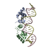

 PDBj
PDBj


 Assembly
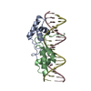
 Sample preparation
 Processing
 NMRPipe