 Movie
Movie Controller

[English] 日本語
 Yorodumi
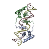
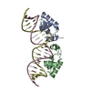
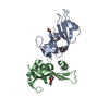
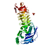
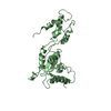
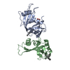
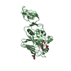
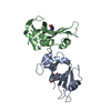
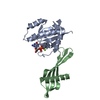
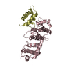
- PDB-1l1m: SOLUTION STRUCTURE OF A DIMER OF LAC REPRESSOR DNA-BINDING DOMAIN... -
+ Open data

- Basic information
| Entry | Database: PDB / ID: 1l1m | ||||||
|---|---|---|---|---|---|---|---|
| Title | SOLUTION STRUCTURE OF A DIMER OF LAC REPRESSOR DNA-BINDING DOMAIN COMPLEXED TO ITS NATURAL OPERATOR O1 | ||||||
 Components Components |
| ||||||
 Keywords Keywords | TRANSCRIPTION REGULATOR/DNA / TRANSCRIPTION REGULATION / LAC OPERON / LAC REPRESSOR / NATURAL LAC OPERATOR / ASYMMETRIC DNA-BINDING / HTH / TRANSCRIPTION REGULATOR-DNA COMPLEX | ||||||
| Function / homology |  Function and homology information Function and homology informationDNA-binding transcription repressor activity / cis-regulatory region sequence-specific DNA binding / transcription cis-regulatory region binding / DNA-binding transcription factor activity / negative regulation of DNA-templated transcription / regulation of DNA-templated transcription / identical protein binding / cytosol Similarity search - Function | ||||||
| Biological species |  | ||||||
| Method | SOLUTION NMR / SIMULATED ANNEALING, RESTRAINED MDR | ||||||
 Authors Authors | Kalodimos, C.G. / Bonvin, A.M.J.J. / Salinas, R.K. / Wechselberger, R. / Boelens, R. / Kaptein, R. | ||||||
 Citation Citation | Journal: EMBO J. / Year: 2002 Title: Plasticity in protein-DNA recognition: lac repressor interacts with its natural operator 01 through alternative conformations of its DNA-binding domain. Authors: Kalodimos, C.G. / Bonvin, A.M. / Salinas, R.K. / Wechselberger, R. / Boelens, R. / Kaptein, R. #1: Journal: Proc.Natl.Acad.Sci.USA / Year: 2001Title: Strong DNA binding by covalently linked dimeric lac headpiece: evidence for the crucial role of the hinge helices Authors: Kalodimos, C.G. / Folkers, G. / Boelens, R. / Kaptein, R. | ||||||
| History |
|
- Structure visualization
| Structure viewer | Molecule: MolmilJmol/JSmol |
|---|
- Downloads & links
-Download
| PDBx/mmCIF format | 1l1m.cif.gz | 1.4 MB | Display | PDBx/mmCIF format |
|---|---|---|---|---|
| PDB format | pdb1l1m.ent.gz | 1.2 MB | Display | PDB format |
| PDBx/mmJSON format | 1l1m.json.gz | Tree view | PDBx/mmJSON format | |
| Others |  Other downloads Other downloads |
-Validation report
| Arichive directory | https://data.pdbj.org/pub/pdb/validation_reports/l1/1l1mftp://data.pdbj.org/pub/pdb/validation_reports/l1/1l1m | HTTPS FTP |
|---|
-Related structure data
| Similar structure data |
|---|
-Links
 PDBj
PDBj


- Assembly
| Deposited unit | 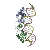
| |||||||||
|---|---|---|---|---|---|---|---|---|---|---|
| 1 |
| |||||||||
| NMR ensembles |
|
-Components
| #1: DNA chain | Mass: 7143.645 Da / Num. of mol.: 1 / Source method: obtained synthetically Details: Lac operator O1; this is the wild-type operator sequence of the lac operon in E. coli. | ||
|---|---|---|---|
| #2: DNA chain | Mass: 6974.534 Da / Num. of mol.: 1 / Source method: obtained synthetically Details: Lac operator O1; this is the wild-type operator sequence of the lac operon in E. coli. | ||
| #3: Protein | Mass: 6822.755 Da / Num. of mol.: 2 / Fragment: N-terminal DNA-binding domain, Residues 1-62 / Mutation: V52C Source method: isolated from a genetically manipulated source Source: (gene. exp.) Has protein modification | Y | |
-Experimental details
-Experiment
| Experiment | Method: SOLUTION NMR | ||||||||||||||||
|---|---|---|---|---|---|---|---|---|---|---|---|---|---|---|---|---|---|
| NMR experiment |
| ||||||||||||||||
| NMR details | Text: This structure was determined using standard 2D and 3D homo- and heteronuclearnuclear techniques. 13C-15N labeled protein was used and unlabeled nucleotide. IN ADDITION ISOTOPE FILTER ...Text: This structure was determined using standard 2D and 3D homo- and heteronuclearnuclear techniques. 13C-15N labeled protein was used and unlabeled nucleotide. IN ADDITION ISOTOPE FILTER EXPERIMENTS WERE APPLIED TO OBTAIN ADDITIONAL ASSIGNMENTS AND TO ASSIGN INTER-MOLECULAR NOES. FOR FURTHER DETAILS SEE THE REFERENCE DESCRIBING THE STRUCTURES. |
- Sample preparation
| Details | Contents: 2mM Lac-HP62-V52C U-15N,13C, 10mM KPi, 20mM KCl, 90% H2O, 10% D2O Solvent system: 90% H2O/10% D2O |
|---|---|
| Sample conditions | Ionic strength: 10mM KPi, 20mM KCl / pH: 6 / Pressure: ambient / Temperature: 315 K |
| Crystal grow | *PLUS Method: other / Details: NMR |
-NMR measurement
| Radiation | Protocol: SINGLE WAVELENGTH / Monochromatic (M) / Laue (L): M | |||||||||||||||
|---|---|---|---|---|---|---|---|---|---|---|---|---|---|---|---|---|
| Radiation wavelength | Relative weight: 1 | |||||||||||||||
| NMR spectrometer |
|
- Processing
| NMR software |
| ||||||||||||||||||||||||
|---|---|---|---|---|---|---|---|---|---|---|---|---|---|---|---|---|---|---|---|---|---|---|---|---|---|
| Refinement | Method: SIMULATED ANNEALING, RESTRAINED MDR / Software ordinal: 1 Details: The structure of the complex was calculated as follows. First the structure of the dimeric lac HP62-V52C was calculated using only protein NMR restraints. The 50 best structures were ...Details: The structure of the complex was calculated as follows. First the structure of the dimeric lac HP62-V52C was calculated using only protein NMR restraints. The 50 best structures were selected and docked onto the natural lac operator B-DNA using simulated annealing. Distance and planarity restraints for the DNA were incorporated in order to keep DNA close to B-DNA but allowing a bend necessary to accommodate the two headpiece molecules on the DNA. | ||||||||||||||||||||||||
| NMR representative | Selection criteria: closest to the average | ||||||||||||||||||||||||
| NMR ensemble | Conformer selection criteria: structures with the lowest energy Conformers calculated total number: 100 / Conformers submitted total number: 20 |
 NMRPipe