 Movie
Movie Controller


+ Open data

- Basic information
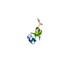
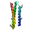
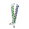
| Entry | Database: PDB / ID: 1okh | ||||||
|---|---|---|---|---|---|---|---|
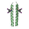
| Title | Viscotoxin A3 from Viscum album L. | ||||||
 Components Components | VISCOTOXIN A3 | ||||||
 Keywords Keywords | TOXIN / THIONIN / PLANT DEFENSE | ||||||
| Function / homology |  Function and homology information Function and homology information | ||||||
| Biological species |  VISCUM ALBUM (European mistletoe) VISCUM ALBUM (European mistletoe) | ||||||
| Method |  X-RAY DIFFRACTION / SYNCHROTRON / SAD / Resolution: 1.75 Å X-RAY DIFFRACTION / SYNCHROTRON / SAD / Resolution: 1.75 Å | ||||||
 Authors Authors | Debreczeni, J.E. / Girmann, B. / Zeeck, A. / Sheldrick, G.M. | ||||||
 Citation Citation | Journal: Acta Crystallogr.,Sect.D / Year: 2003 Title: Structure of Viscotoxin A3: Disulfide Location from Weak Sad Data Authors: Debreczeni, J.E. / Girmann, B. / Zeeck, A. / Kratzner, R. / Sheldrick, G.M. | ||||||
| History |
|
- Structure visualization
| Structure viewer | Molecule: MolmilJmol/JSmol |
|---|
- Downloads & links
-Download
| PDBx/mmCIF format | 1okh.cif.gz | 29.8 KB | Display | PDBx/mmCIF format |
|---|---|---|---|---|
| PDB format | pdb1okh.ent.gz | 20.3 KB | Display | PDB format |
| PDBx/mmJSON format | 1okh.json.gz | Tree view | PDBx/mmJSON format | |
| Others |  Other downloads Other downloads |
-Validation report
| Arichive directory | https://data.pdbj.org/pub/pdb/validation_reports/ok/1okhftp://data.pdbj.org/pub/pdb/validation_reports/ok/1okh | HTTPS FTP |
|---|
-Related structure data
| Similar structure data |
|---|

-Links
 PDBj
PDBj
- Assembly
| Deposited unit | 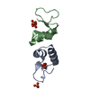
| |||||||||||||||
|---|---|---|---|---|---|---|---|---|---|---|---|---|---|---|---|---|
| 1 |
| |||||||||||||||
| Unit cell |
| |||||||||||||||
| Components on special symmetry positions |
| |||||||||||||||
| Noncrystallographic symmetry (NCS) | NCS oper: (Code: given Matrix: (0.19986, -0.97837, -0.05339), Vector: |
-Components
| #1: Protein/peptide | Mass: 4842.626 Da / Num. of mol.: 2 / Fragment: VISCOTOXIN A3 CHAIN, RESIDUES 27-72 / Source method: isolated from a natural source / Source: (natural) VISCUM ALBUM (European mistletoe) / Organ: LEAVES, STEMS / References: UniProt: P01538#2: Chemical |   Mass: 94.971 Da / Num. of mol.: 2 / Source method: obtained synthetically / Formula: PO4 Mass: 94.971 Da / Num. of mol.: 2 / Source method: obtained synthetically / Formula: PO4#3: Chemical | ChemComp-SO4 / |   Mass: 96.063 Da / Num. of mol.: 1 / Source method: obtained synthetically / Formula: SO4 Mass: 96.063 Da / Num. of mol.: 1 / Source method: obtained synthetically / Formula: SO4#4: Water | ChemComp-HOH / |  Mass: 18.015 Da / Num. of mol.: 94 / Source method: isolated from a natural source / Formula: H2O Mass: 18.015 Da / Num. of mol.: 94 / Source method: isolated from a natural source / Formula: H2OHas protein modification | Y | |
|---|
-Experimental details
-Experiment
| Experiment | Method: X-RAY DIFFRACTION / Number of used crystals: 1 |
|---|
- Sample preparation
| Crystal | Density Matthews: 1.79 Å3/Da / Density % sol: 31.5 % Description: DATA COLLECTED IN-HOUSE. PHASED USING IN-HOUSE SULFUR- SAD DATA | ||||||||||||||||||||||||||||||||||||||||||
|---|---|---|---|---|---|---|---|---|---|---|---|---|---|---|---|---|---|---|---|---|---|---|---|---|---|---|---|---|---|---|---|---|---|---|---|---|---|---|---|---|---|---|---|
| Crystal grow | pH: 6.5 Details: 0.15M AM2SO4, 0.05M CACOD. PH=6.5, 30% PEG8000, 15MM HGCL2, pH 6.50 | ||||||||||||||||||||||||||||||||||||||||||
| Crystal grow | *PLUS pH: 6.5 / Method: vapor diffusion, hanging drop | ||||||||||||||||||||||||||||||||||||||||||
| Components of the solutions | *PLUS
|
-Data collection
| Diffraction | Mean temperature: 100 K |
|---|---|
| Diffraction source | Source: SYNCHROTRON / Site: EMBL/DESY, HAMBURG  / Beamline: X13 / Wavelength: 0.98 / Beamline: X13 / Wavelength: 0.98 |
| Detector | Type: MARRESEARCH / Detector: IMAGE PLATE / Date: Apr 15, 2002 |
| Radiation | Protocol: SINGLE WAVELENGTH / Monochromatic (M) / Laue (L): M / Scattering type: x-ray |
| Radiation wavelength | Wavelength: 0.98 Å / Relative weight: 1 |
| Reflection | Resolution: 1.75→68.59 Å / Num. obs: 8938 / % possible obs: 100 % / Redundancy: 5.25 % / Rmerge(I) obs: 0.0892 / Net I/σ(I): 14.31 |
| Reflection shell | Resolution: 1.75→1.85 Å / Redundancy: 4.42 % / Rmerge(I) obs: 0.3866 / Mean I/σ(I) obs: 3.89 / % possible all: 99.7 |
| Reflection | *PLUS Highest resolution: 1.75 Å / Redundancy: 5.3 % / Num. measured all: 50787 / Rmerge(I) obs: 0.089 |
| Reflection shell | *PLUS % possible obs: 99.7 % / Redundancy: 4.4 % / Rmerge(I) obs: 0.387 / Mean I/σ(I) obs: 3.9 |
- Processing
| Software |
| |||||||||||||||||||||||||||||||||
|---|---|---|---|---|---|---|---|---|---|---|---|---|---|---|---|---|---|---|---|---|---|---|---|---|---|---|---|---|---|---|---|---|---|---|
| Refinement | Method to determine structure: SAD / Resolution: 1.75→20 Å / Num. parameters: 3115 / Num. restraintsaints: 2935 / Cross valid method: FREE R-VALUE / σ(F): 0 / Stereochemistry target values: ENGH AND HUBER
| |||||||||||||||||||||||||||||||||
| Refine analyze | Num. disordered residues: 1 / Occupancy sum hydrogen: 0 / Occupancy sum non hydrogen: 780 | |||||||||||||||||||||||||||||||||
| Refinement step | Cycle: LAST / Resolution: 1.75→20 Å
| |||||||||||||||||||||||||||||||||
| Refine LS restraints |
| |||||||||||||||||||||||||||||||||
| Software | *PLUS Name: SHELXL / Version: 97 / Classification: refinement | |||||||||||||||||||||||||||||||||
| Refinement | *PLUS Rfactor Rfree: 0.239 / Rfactor Rwork: 0.167 | |||||||||||||||||||||||||||||||||
| Solvent computation | *PLUS | |||||||||||||||||||||||||||||||||
| Displacement parameters | *PLUS | |||||||||||||||||||||||||||||||||
| Refine LS restraints | *PLUS
|
