 Movie
Movie Controller

[English] 日本語
 Yorodumi
- PDB-1o8s: Structure of CsCBM6-3 from Clostridium stercorarium in complex wi... -
+ Open data

- Basic information
| Entry | Database: PDB / ID: 1o8s | |||||||||
|---|---|---|---|---|---|---|---|---|---|---|
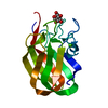
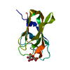
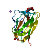
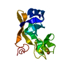
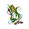
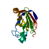
| Title | Structure of CsCBM6-3 from Clostridium stercorarium in complex with cellobiose | |||||||||
 Components Components | PUTATIVE ENDO-XYLANASE | |||||||||
 Keywords Keywords |  HYDROLASE / CARBOHYDRATE-BINDING MODULE / XYLAN / CELLULOSE / BETA- SANDWICH / GLYCOSIDASE / XYLAN DEGRADATION HYDROLASE / CARBOHYDRATE-BINDING MODULE / XYLAN / CELLULOSE / BETA- SANDWICH / GLYCOSIDASE / XYLAN DEGRADATION | |||||||||
| Function / homology |  Function and homology informationendo-1,4-beta-xylanase activity / endo-1,4-beta-xylanase / xylan catabolic process / cellulose catabolic process / carbohydrate binding / extracellular region / metal ion binding Function and homology informationendo-1,4-beta-xylanase activity / endo-1,4-beta-xylanase / xylan catabolic process / cellulose catabolic process / carbohydrate binding / extracellular region / metal ion bindingSimilarity search - Function | |||||||||
| Biological species |  CLOSTRIDIUM STERCORARIUM (bacteria) CLOSTRIDIUM STERCORARIUM (bacteria) | |||||||||
| Method | X-RAY DIFFRACTION / SYNCHROTRON / MAD / Resolution: 1.15 Å | |||||||||
 Authors Authors | Boraston, A.B. / Notenboom, V. / Warren, R.A.J. / Kilbrun, D.G. / Rose, D.R. / Davies, G.J. | |||||||||
 Citation Citation | Journal: J.Mol.Biol. / Year: 2003 Title: Structure and Ligand Binding of Carbohydrate-Binding Module Cscbm6-3 Reveals Similarities with Fucose-Specific Lectins and Galactose-Binding Domains Authors: Boraston, A.B. / Notenboom, V. / Warren, R.A.J. / Kilburn, D.G. / Rose, D.R. / Davies, G.J. | |||||||||
| History |
| |||||||||
| Remark 700 | SHEET THE SHEET STRUCTURE OF THIS MOLECULE IS BIFURCATED. IN ORDER TO REPRESENT THIS FEATURE IN ... SHEET THE SHEET STRUCTURE OF THIS MOLECULE IS BIFURCATED. IN ORDER TO REPRESENT THIS FEATURE IN THE SHEET RECORDS BELOW, TWO SHEETS ARE DEFINED. |
- Structure visualization
| Structure viewer | Molecule: MolmilJmol/JSmol |
|---|
- Downloads & links
-Download
| PDBx/mmCIF format | 1o8s.cif.gz | 75.9 KB | Display | PDBx/mmCIF format |
|---|---|---|---|---|
| PDB format | pdb1o8s.ent.gz | 53.8 KB | Display | PDB format |
| PDBx/mmJSON format | 1o8s.json.gz | Tree view | PDBx/mmJSON format | |
| Others |  Other downloads Other downloads |
-Validation report
| Arichive directory | https://data.pdbj.org/pub/pdb/validation_reports/o8/1o8sftp://data.pdbj.org/pub/pdb/validation_reports/o8/1o8s | HTTPS FTP |
|---|
-Related structure data
| Related structure data |  1naeC  1o8pC  1od3C 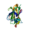 1gmmS C: citing same article ( S: Starting model for refinement |
|---|---|
| Similar structure data |


-Links
 PDBj
PDBj
- Assembly
| Deposited unit | 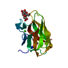
| ||||||||
|---|---|---|---|---|---|---|---|---|---|
| 1 |
| ||||||||
| Unit cell |
|
-Components
| #1: Protein | Mass: 17405.947 Da / Num. of mol.: 1 / Fragment: CARBOHYDRATE-BINDING DOMAIN, RESIDUES 273-417 Source method: isolated from a genetically manipulated source Source: (gene. exp.) CLOSTRIDIUM STERCORARIUM (bacteria) / Strain: NCIB11745 / Plasmid: PET28A / Production host: ESCHERICHIA COLI (E. coli) / Strain (production host): BL21(DE3)References: UniProt: Q93AQ5, UniProt: Q8GJ44*PLUS, endo-1,4-beta-xylanase |
|---|---|
| #2: Polysaccharide | beta-D-glucopyranose-(1-4)-beta-D-glucopyranose / beta-cellobiose  , Oligosaccharide / Class: Metabolism / Mass: 342.297 Da / Num. of mol.: 1 , Oligosaccharide / Class: Metabolism / Mass: 342.297 Da / Num. of mol.: 1Source method: isolated from a genetically manipulated source Details: oligosaccharide / References: beta-cellobiose |
| #3: Chemical | ChemComp-CA /   Mass: 40.078 Da / Num. of mol.: 1 / Source method: obtained synthetically / Formula: Ca Mass: 40.078 Da / Num. of mol.: 1 / Source method: obtained synthetically / Formula: Ca |
| #4: Water | ChemComp-HOH / Water Mass: 18.015 Da / Num. of mol.: 205 / Source method: isolated from a natural source / Formula: H2O Mass: 18.015 Da / Num. of mol.: 205 / Source method: isolated from a natural source / Formula: H2O |
| Sequence details | C-TERMINAL MODULE |
-Experimental details
-Experiment
| Experiment | Method: X-RAY DIFFRACTION / Number of used crystals: 1 |
|---|
- Sample preparation
| Crystal | Density Matthews: 1.75 Å3/Da / Density % sol: 29.61 % | ||||||||||||||||||||||||||||||
|---|---|---|---|---|---|---|---|---|---|---|---|---|---|---|---|---|---|---|---|---|---|---|---|---|---|---|---|---|---|---|---|
| Crystal grow | pH: 4.6 / Details: pH 4.60 | ||||||||||||||||||||||||||||||
| Crystal grow | *PLUS Method: vapor diffusion, hanging drop | ||||||||||||||||||||||||||||||
| Components of the solutions | *PLUS
|
-Data collection
| Diffraction | Mean temperature: 100 K |
|---|---|
| Diffraction source | Source: SYNCHROTRON / Site: ESRF  / Beamline: ID29 / Wavelength: 0.9795 / Beamline: ID29 / Wavelength: 0.9795 |
| Detector | Type: ADSC CCD / Detector: CCD |
| Radiation | Protocol: SINGLE WAVELENGTH / Monochromatic (M) / Laue (L): M / Scattering type: x-ray |
| Radiation wavelength | Wavelength: 0.9795 Å / Relative weight: 1 |
| Reflection | Resolution: 1.15→40 Å / Num. obs: 66726 / % possible obs: 97.8 % / Redundancy: 3.7 % / Rmerge(I) obs: 0.072 / Net I/σ(I): 15.6 |
| Reflection shell | Resolution: 1.15→1.21 Å / Redundancy: 1.6 % / Rmerge(I) obs: 0.293 / Mean I/σ(I) obs: 2.8 / % possible all: 15.3 |
| Reflection | *PLUS Highest resolution: 1.35 Å / Lowest resolution: 40 Å |
| Reflection shell | *PLUS Highest resolution: 1.35 Å / Lowest resolution: 1.42 Å / % possible obs: 86.5 % / Redundancy: 3.2 % / Rmerge(I) obs: 0.162 / Mean I/σ(I) obs: 6.9 |
- Processing
| Software |
| ||||||||||||||||||||||||||||||||||||||||||||||||||||||||||||||||||||||||||||||||||||||||||||||||||||||||||||||||||||||||||||||||||||||||||||||||||||||||||||||||||||||||||||||||||||||
|---|---|---|---|---|---|---|---|---|---|---|---|---|---|---|---|---|---|---|---|---|---|---|---|---|---|---|---|---|---|---|---|---|---|---|---|---|---|---|---|---|---|---|---|---|---|---|---|---|---|---|---|---|---|---|---|---|---|---|---|---|---|---|---|---|---|---|---|---|---|---|---|---|---|---|---|---|---|---|---|---|---|---|---|---|---|---|---|---|---|---|---|---|---|---|---|---|---|---|---|---|---|---|---|---|---|---|---|---|---|---|---|---|---|---|---|---|---|---|---|---|---|---|---|---|---|---|---|---|---|---|---|---|---|---|---|---|---|---|---|---|---|---|---|---|---|---|---|---|---|---|---|---|---|---|---|---|---|---|---|---|---|---|---|---|---|---|---|---|---|---|---|---|---|---|---|---|---|---|---|---|---|---|---|
| Refinement | Method to determine structure: MAD Starting model: PDB ENTRY 1GMM Resolution: 1.15→40.49 Å / Cor.coef. Fo:Fc: 0.978 / Cor.coef. Fo:Fc free: 0.968 / SU B: 0.492 / SU ML: 0.022 / Cross valid method: THROUGHOUT / ESU R: 0.039 / ESU R Free: 0.038 / Stereochemistry target values: MAXIMUM LIKELIHOOD / Details: HYDROGENS HAVE BEEN ADDED IN THE RIDING POSITIONS
| ||||||||||||||||||||||||||||||||||||||||||||||||||||||||||||||||||||||||||||||||||||||||||||||||||||||||||||||||||||||||||||||||||||||||||||||||||||||||||||||||||||||||||||||||||||||
| Solvent computation | Ion probe radii: 0.8 Å / Shrinkage radii: 0.8 Å / VDW probe radii: 1.4 Å / Solvent model: BABINET MODEL WITH MASK | ||||||||||||||||||||||||||||||||||||||||||||||||||||||||||||||||||||||||||||||||||||||||||||||||||||||||||||||||||||||||||||||||||||||||||||||||||||||||||||||||||||||||||||||||||||||
| Displacement parameters | Biso mean: 8.3 Å2
| ||||||||||||||||||||||||||||||||||||||||||||||||||||||||||||||||||||||||||||||||||||||||||||||||||||||||||||||||||||||||||||||||||||||||||||||||||||||||||||||||||||||||||||||||||||||
| Refinement step | Cycle: LAST / Resolution: 1.15→40.49 Å
| ||||||||||||||||||||||||||||||||||||||||||||||||||||||||||||||||||||||||||||||||||||||||||||||||||||||||||||||||||||||||||||||||||||||||||||||||||||||||||||||||||||||||||||||||||||||
| Refine LS restraints |
|