 Movie
Movie Controller

[English] 日本語
 Yorodumi
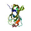
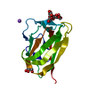
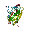
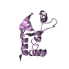
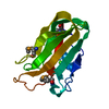
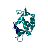
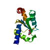
- PDB-1nae: Structure of CsCBM6-3 from Clostridium stercorarium in complex wi... -
+ Open data

- Basic information
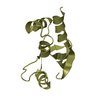
| Entry | Database: PDB / ID: 1nae | |||||||||
|---|---|---|---|---|---|---|---|---|---|---|
| Title | Structure of CsCBM6-3 from Clostridium stercorarium in complex with xylotriose | |||||||||
 Components Components | putative xylanase | |||||||||
 Keywords Keywords | HYDROLASE / carbohydrate-binding module / beta-sandwich / xylan / xylooligosaccharide | |||||||||
| Function / homology |  Function and homology information Function and homology informationendo-1,4-beta-xylanase / endo-1,4-beta-xylanase activity / xylan catabolic process / cellulose catabolic process / carbohydrate binding / extracellular region / metal ion binding Similarity search - Function | |||||||||
| Biological species |  Clostridium stercorarium (bacteria) Clostridium stercorarium (bacteria) | |||||||||
| Method |  X-RAY DIFFRACTION / MOLECULAR REPLACEMENT / Resolution: 2.05 Å X-RAY DIFFRACTION / MOLECULAR REPLACEMENT / Resolution: 2.05 Å | |||||||||
 Authors Authors | Boraston, A.B. / Notenboom, V. / Warren, R.A.J. / Kilburn, D.G. / Rose, D.R. / Davies, G. | |||||||||
 Citation Citation | Journal: J.Mol.Biol. / Year: 2003 Title: Structure and ligand binding of carbohydrate-binding module CsCBM6-3 reveals similarities with fucose-specific lectins and "galactose-binding" domains Authors: Boraston, A.B. / Notenboom, V. / Warren, R.A.J. / Kilburn, D.G. / Rose, D.R. / Davies, G. | |||||||||
| History |
|
- Structure visualization
| Structure viewer | Molecule: MolmilJmol/JSmol |
|---|
- Downloads & links
-Download
| PDBx/mmCIF format | 1nae.cif.gz | 45 KB | Display | PDBx/mmCIF format |
|---|---|---|---|---|
| PDB format | pdb1nae.ent.gz | 28.9 KB | Display | PDB format |
| PDBx/mmJSON format | 1nae.json.gz | Tree view | PDBx/mmJSON format | |
| Others |  Other downloads Other downloads |
-Validation report
| Arichive directory | https://data.pdbj.org/pub/pdb/validation_reports/na/1naeftp://data.pdbj.org/pub/pdb/validation_reports/na/1nae | HTTPS FTP |
|---|
-Related structure data
| Related structure data |  1o8pC  1o8sC  1od3C 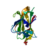 1gmmS S: Starting model for refinement C: citing same article ( |
|---|---|
| Similar structure data |


-Links
 PDBj
PDBj
- Assembly
| Deposited unit | 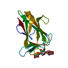
| ||||||||
|---|---|---|---|---|---|---|---|---|---|
| 1 |
| ||||||||
| Unit cell |
|
-Components
| #1: Protein | Mass: 17405.947 Da / Num. of mol.: 1 / Fragment: C-terminal carbohydrate-binding module Source method: isolated from a genetically manipulated source Source: (gene. exp.) Clostridium stercorarium (bacteria) / Gene: putative xylanase / Plasmid: pET28a / Species (production host): Escherichia coli / Production host: |
|---|---|
| #2: Polysaccharide | beta-D-xylopyranose-(1-4)-beta-D-xylopyranose-(1-4)-beta-D-xylopyranose / 4beta-beta-xylotriose  Source method: isolated from a genetically manipulated source Details: oligosaccharide / References: 4beta-beta-xylotriose |
| #3: Chemical | ChemComp-CA /   Mass: 40.078 Da / Num. of mol.: 1 / Source method: obtained synthetically / Formula: Ca Mass: 40.078 Da / Num. of mol.: 1 / Source method: obtained synthetically / Formula: Ca |
| #4: Water | ChemComp-HOH /  Mass: 18.015 Da / Num. of mol.: 178 / Source method: isolated from a natural source / Formula: H2O Mass: 18.015 Da / Num. of mol.: 178 / Source method: isolated from a natural source / Formula: H2O |
-Experimental details
-Experiment
| Experiment | Method: X-RAY DIFFRACTION / Number of used crystals: 1 |
|---|
- Sample preparation
| Crystal | Density Matthews: 1.63 Å3/Da / Density % sol: 24.09 % | ||||||||||||||||||||||||||||||
|---|---|---|---|---|---|---|---|---|---|---|---|---|---|---|---|---|---|---|---|---|---|---|---|---|---|---|---|---|---|---|---|
| Crystal grow | Temperature: 291 K / Method: vapor diffusion, hanging drop / pH: 4.6 Details: 26%PEG 4K, 0.1 M sodium acetate, pH 4.6, 0.2 M ammonium sulfate, VAPOR DIFFUSION, HANGING DROP, temperature 291K | ||||||||||||||||||||||||||||||
| Crystal grow | *PLUS | ||||||||||||||||||||||||||||||
| Components of the solutions | *PLUS
|
-Data collection
| Diffraction | Mean temperature: 100 K |
|---|---|
| Diffraction source | Source: ROTATING ANODE / Type: RIGAKU / Wavelength: 1.54 Å |
| Detector | Type: MARRESEARCH / Detector: IMAGE PLATE / Date: Jul 12, 2002 / Details: long mirror focused |
| Radiation | Protocol: SINGLE WAVELENGTH / Monochromatic (M) / Laue (L): M / Scattering type: x-ray |
| Radiation wavelength | Wavelength: 1.54 Å / Relative weight: 1 |
| Reflection | Resolution: 2.05→36.04 Å / Num. all: 6680 / Num. obs: 6382 / Redundancy: 2.3 % / Biso Wilson estimate: 20.33 Å2 / Rmerge(I) obs: 0.07 / Rsym value: 0.07 / Net I/σ(I): 12.4 |
| Reflection shell | Resolution: 2.05→2.12 Å / Redundancy: 2.2 % / Rmerge(I) obs: 0.186 / Mean I/σ(I) obs: 5.4 / Num. unique all: 649 / Rsym value: 0.186 |
| Reflection | *PLUS Highest resolution: 2.25 Å / Lowest resolution: 40 Å / % possible obs: 94.6 % / Redundancy: 8.5 % / Rmerge(I) obs: 0.046 |
| Reflection shell | *PLUS Highest resolution: 2.25 Å / Lowest resolution: 2.36 Å / % possible obs: 72.1 % / Redundancy: 5 % / Rmerge(I) obs: 0.141 / Mean I/σ(I) obs: 6.6 |
- Processing
| Software |
| |||||||||||||||||||||||||||||||||||||||||||||||||||||||||||||||||||||||||||
|---|---|---|---|---|---|---|---|---|---|---|---|---|---|---|---|---|---|---|---|---|---|---|---|---|---|---|---|---|---|---|---|---|---|---|---|---|---|---|---|---|---|---|---|---|---|---|---|---|---|---|---|---|---|---|---|---|---|---|---|---|---|---|---|---|---|---|---|---|---|---|---|---|---|---|---|---|
| Refinement | Method to determine structure: MOLECULAR REPLACEMENT Starting model: 1gmm Resolution: 2.05→36.04 Å / Cor.coef. Fo:Fc: 0.97 / Cor.coef. Fo:Fc free: 0.946 / SU B: 4.63 / SU ML: 0.126 / Isotropic thermal model: Isotropic / Cross valid method: THROUGHOUT / ESU R: 0.334 / ESU R Free: 0.194
| |||||||||||||||||||||||||||||||||||||||||||||||||||||||||||||||||||||||||||
| Solvent computation | Ion probe radii: 0.8 Å / Shrinkage radii: 0.8 Å / VDW probe radii: 1.4 Å / Solvent model: BABINET MODEL WITH MASK | |||||||||||||||||||||||||||||||||||||||||||||||||||||||||||||||||||||||||||
| Displacement parameters | Biso mean: 19.907 Å2
| |||||||||||||||||||||||||||||||||||||||||||||||||||||||||||||||||||||||||||
| Refinement step | Cycle: LAST / Resolution: 2.05→36.04 Å
| |||||||||||||||||||||||||||||||||||||||||||||||||||||||||||||||||||||||||||
| Refine LS restraints |
| |||||||||||||||||||||||||||||||||||||||||||||||||||||||||||||||||||||||||||
| LS refinement shell | Resolution: 2.05→2.103 Å / Total num. of bins used: 20
| |||||||||||||||||||||||||||||||||||||||||||||||||||||||||||||||||||||||||||
| Refinement | *PLUS Highest resolution: 2.25 Å / Lowest resolution: 40 Å / Rfactor Rfree: 0.203 / Rfactor Rwork: 0.161 | |||||||||||||||||||||||||||||||||||||||||||||||||||||||||||||||||||||||||||
| Solvent computation | *PLUS | |||||||||||||||||||||||||||||||||||||||||||||||||||||||||||||||||||||||||||
| Displacement parameters | *PLUS | |||||||||||||||||||||||||||||||||||||||||||||||||||||||||||||||||||||||||||
| Refine LS restraints | *PLUS
| |||||||||||||||||||||||||||||||||||||||||||||||||||||||||||||||||||||||||||
| LS refinement shell | *PLUS Highest resolution: 2.25 Å / Lowest resolution: 2.36 Å |
