 Movie
Movie Controller

[English] 日本語
 Yorodumi
- PDB-1nrr: Crystallographic structures of Thrombin complexed with Thrombin r... -
+ Open data

- Basic information
| Entry | Database: PDB / ID: 1nrr | ||||||
|---|---|---|---|---|---|---|---|
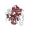
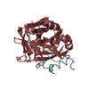
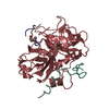
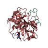
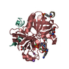
| Title | Crystallographic structures of Thrombin complexed with Thrombin receptor peptides: Existence of expected and novel binding modes | ||||||
 Components Components |
| ||||||
 Keywords Keywords | Hydrolase/HYDROLASE INHIBITOR / Hydrolase-Hydrolase Inhibitor complex /  BLOOD CLOTTING BLOOD CLOTTING | ||||||
| Function / homology |  Function and homology information Function and homology informationnegative regulation of renin secretion into blood stream / dendritic cell homeostasis / negative regulation of glomerular filtration / thrombin-activated receptor activity / establishment of synaptic specificity at neuromuscular junction / platelet dense tubular network / cell-cell junction maintenance / regulation of interleukin-1 beta production / connective tissue replacement involved in inflammatory response wound healing / platelet dense granule organization ...negative regulation of renin secretion into blood stream / dendritic cell homeostasis / negative regulation of glomerular filtration / thrombin-activated receptor activity / establishment of synaptic specificity at neuromuscular junction / platelet dense tubular network / cell-cell junction maintenance / regulation of interleukin-1 beta production / connective tissue replacement involved in inflammatory response wound healing / platelet dense granule organization / trans-synaptic signaling by endocannabinoid, modulating synaptic transmission / regulation of sensory perception of pain / positive regulation of smooth muscle contraction / protein kinase C-activating G protein-coupled receptor signaling pathway / positive regulation of calcium ion transport / positive regulation of Rho protein signal transduction / positive regulation of lipid kinase activity / positive regulation of phospholipase C-activating G protein-coupled receptor signaling pathway / cytolysis by host of symbiont cells / thrombospondin receptor activity / Defective factor XII causes hereditary angioedema / thrombin / neutrophil-mediated killing of gram-negative bacterium / regulation of blood coagulation / ligand-gated ion channel signaling pathway / Defective F8 cleavage by thrombin / Platelet Aggregation (Plug Formation) / positive regulation of cysteine-type endopeptidase activity involved in apoptotic process / negative regulation of platelet activation / negative regulation of astrocyte differentiation / G-protein alpha-subunit binding / anatomical structure morphogenesis / positive regulation of collagen biosynthetic process / negative regulation of cytokine production involved in inflammatory response / positive regulation of blood coagulation / negative regulation of fibrinolysis / Gamma-carboxylation of protein precursors / Transport of gamma-carboxylated protein precursors from the endoplasmic reticulum to the Golgi apparatus / Common Pathway of Fibrin Clot Formation / Removal of aminoterminal propeptides from gamma-carboxylated proteins / homeostasis of number of cells within a tissue / release of sequestered calcium ion into cytosol / positive regulation of vasoconstriction / fibrinolysis / regulation of cytosolic calcium ion concentration / Intrinsic Pathway of Fibrin Clot Formation / Peptide ligand-binding receptors / positive regulation of release of sequestered calcium ion into cytosol / Regulation of Complement cascade / caveola / G protein-coupled receptor activity / positive regulation of interleukin-8 production / acute-phase response / Cell surface interactions at the vascular wall / lipopolysaccharide binding / negative regulation of proteolysis / positive regulation of receptor signaling pathway via JAK-STAT / growth factor activity / neuromuscular junction / positive regulation of insulin secretion / platelet activation / positive regulation of GTPase activity / response to wounding / positive regulation of interleukin-6 production / Golgi lumen / positive regulation of protein localization to nucleus / Regulation of Insulin-like Growth Factor (IGF) transport and uptake by Insulin-like Growth Factor Binding Proteins (IGFBPs) / G-protein beta-subunit binding / activation of cysteine-type endopeptidase activity involved in apoptotic process / positive regulation of reactive oxygen species metabolic process / blood coagulation / antimicrobial humoral immune response mediated by antimicrobial peptide / Thrombin signalling through proteinase activated receptors (PARs) / late endosome / heparin binding / phospholipase C-activating G protein-coupled receptor signaling pathway / positive regulation of cytosolic calcium ion concentration / regulation of cell shape / postsynaptic membrane / positive regulation of cell growth / G alpha (q) signalling events / collagen-containing extracellular matrix / blood microparticle / positive regulation of canonical NF-kappaB signal transduction / negative regulation of neuron apoptotic process / response to lipopolysaccharide / positive regulation of MAPK cascade / positive regulation of ERK1 and ERK2 cascade / cell surface receptor signaling pathway / positive regulation of phosphatidylinositol 3-kinase/protein kinase B signal transduction / early endosome / positive regulation of cell migration / inflammatory response / positive regulation of protein phosphorylation / G protein-coupled receptor signaling pathway / endoplasmic reticulum lumen / negative regulation of cell population proliferation / signaling receptor binding / serine-type endopeptidase activity / calcium ion bindingSimilarity search - Function | ||||||
| Biological species |  Homo sapiens (human) Homo sapiens (human) | ||||||
| Method | X-RAY DIFFRACTION / Resolution: 2.4 Å | ||||||
 Authors Authors | Tulinsky, A. / Mathews, I.I. | ||||||
 Citation Citation | Journal: Biochemistry / Year: 1994 Title: Crystallographic structures of thrombin complexed with thrombin receptor peptides: existence of expected and novel binding modes. Authors: Mathews, I.I. / Padmanabhan, K.P. / Ganesh, V. / Tulinsky, A. / Ishii, M. / Chen, J. / Turck, C.W. / Coughlin, S.R. / Fenton 2nd., J.W. #1: Journal: J.Mol.Biol. / Year: 1991Title: Refined Structure of the Hirudin-Thrombin Complex Authors: Rydel, T.J. / Tulinsky, A. / Bode, W. / Huber, R. #2: Journal: J.Mol.Biol. / Year: 1991Title: Structure of the Hirugen and Hirulog 1 Complexes of Alpha-Thrombin Authors: Skrzypczak-Jankun, E. / Carperos, V.E. / Ravichandran, K.G. / Tulinsky, A. / Westbrook, M. / Maraganore, J.M. | ||||||
| History |
|
- Structure visualization
| Structure viewer | Molecule: MolmilJmol/JSmol |
|---|
- Downloads & links
-Download
| PDBx/mmCIF format | 1nrr.cif.gz | 73.5 KB | Display | PDBx/mmCIF format |
|---|---|---|---|---|
| PDB format | pdb1nrr.ent.gz | 57.7 KB | Display | PDB format |
| PDBx/mmJSON format | 1nrr.json.gz | Tree view | PDBx/mmJSON format | |
| Others |  Other downloads Other downloads |
-Validation report
| Arichive directory | https://data.pdbj.org/pub/pdb/validation_reports/nr/1nrrftp://data.pdbj.org/pub/pdb/validation_reports/nr/1nrr | HTTPS FTP |
|---|
-Related structure data


-Links
 PDBj
PDBj


- Assembly
| Deposited unit | 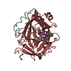
| |||||||||
|---|---|---|---|---|---|---|---|---|---|---|
| 1 |
| |||||||||
| Unit cell |
| |||||||||
| Atom site foot note | 1: CIS PROLINE - PRO H 37 | |||||||||
| Components on special symmetry positions |
|
-Components
| #1: Protein/peptide | / Coagulation factor II Mass: 4096.534 Da / Num. of mol.: 1 / Fragment: RESIDUES 328-363 / Source method: isolated from a natural source / Source: (natural) Homo sapiens (human) / References: UniProt: P00734, thrombin |
|---|---|
| #2: Protein | / Coagulation factor II Mass: 29780.219 Da / Num. of mol.: 1 / Fragment: RESIDUES 364-622 / Source method: isolated from a natural source / Source: (natural) Homo sapiens (human) / References: UniProt: P00734, thrombin |
| #3: Protein/peptide | Mass: 2343.501 Da / Num. of mol.: 1 / Fragment: UNP residues 43-60 / Source method: isolated from a natural source / Source: (natural) Homo sapiens (human) / References: UniProt: P25116 |
| #4: Chemical | ChemComp-0G6 /   Type: peptide-like, Peptide-like / Class: Inhibitor / Mass: 453.986 Da / Num. of mol.: 1 / Source method: obtained synthetically / Formula: C21H34ClN6O3 / References: D-Phe-Pro-Arg-CH2Cl Type: peptide-like, Peptide-like / Class: Inhibitor / Mass: 453.986 Da / Num. of mol.: 1 / Source method: obtained synthetically / Formula: C21H34ClN6O3 / References: D-Phe-Pro-Arg-CH2Cl |
| #5: Water | ChemComp-HOH / Water Mass: 18.015 Da / Num. of mol.: 207 / Source method: isolated from a natural source / Formula: H2O Mass: 18.015 Da / Num. of mol.: 207 / Source method: isolated from a natural source / Formula: H2O |
| Nonpolymer details | THE INHIBITOR IS BOUND TO THE ACTIVE SITE OF THE ENZYME. THE UNBOUND FORM OF THE INHIBITOR IS D-PHE- ...THE INHIBITOR IS BOUND TO THE ACTIVE SITE OF THE ENZYME. THE UNBOUND FORM OF THE INHIBITOR IS D-PHE-PRO-ARG-CHLOROMETH |
| Sequence details | CHYMOTRYPSIN NUMBERING SYSTEM IS USED, BASED ON THE TOPOLOGICAL ALIGNMENT WITH THE STRUCTURE OF ...CHYMOTRYPS |
-Experimental details
-Experiment
| Experiment | Method: X-RAY DIFFRACTION |
|---|
- Sample preparation
| Crystal | Density Matthews: 2.5 Å3/Da / Density % sol: 50.77 % |
|---|---|
| Crystal grow | *PLUS Method: vapor diffusion, hanging drop / PH range low: 8.5 / PH range high: 6.5 |
| Components of the solutions | *PLUS Conc.: 24-30 %(w/v) / Common name: PEG4000 / Details: or PEG8000 |
-Data collection
| Radiation | Scattering type: x-ray |
|---|---|
| Radiation wavelength | Relative weight: 1 |
| Reflection | *PLUS Highest resolution: 2.4 Å / % possible obs: 91 % / Rmerge F obs: 0.035 |
- Processing
| Software | Name: PROLSQ / Classification: refinement | ||||||||||||||||||||||||||||||||||||||||||||||||||||||||||||||||||||||||||||||||||||
|---|---|---|---|---|---|---|---|---|---|---|---|---|---|---|---|---|---|---|---|---|---|---|---|---|---|---|---|---|---|---|---|---|---|---|---|---|---|---|---|---|---|---|---|---|---|---|---|---|---|---|---|---|---|---|---|---|---|---|---|---|---|---|---|---|---|---|---|---|---|---|---|---|---|---|---|---|---|---|---|---|---|---|---|---|---|
| Refinement | Resolution: 2.4→7 Å / σ(F): 2 /
| ||||||||||||||||||||||||||||||||||||||||||||||||||||||||||||||||||||||||||||||||||||
| Refinement step | Cycle: LAST / Resolution: 2.4→7 Å
| ||||||||||||||||||||||||||||||||||||||||||||||||||||||||||||||||||||||||||||||||||||
| Refine LS restraints |
| ||||||||||||||||||||||||||||||||||||||||||||||||||||||||||||||||||||||||||||||||||||
| Refinement | *PLUS Rfactor obs: 0.145 | ||||||||||||||||||||||||||||||||||||||||||||||||||||||||||||||||||||||||||||||||||||
| Solvent computation | *PLUS | ||||||||||||||||||||||||||||||||||||||||||||||||||||||||||||||||||||||||||||||||||||
| Displacement parameters | *PLUS Biso mean: 25 Å2 |
