 Movie
Movie Controller


+ Open data

- Basic information
| Entry | Database: PDB / ID: 1nm3 | ||||||
|---|---|---|---|---|---|---|---|
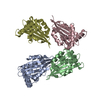
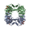
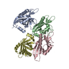
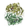
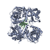
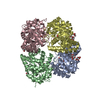
| Title | Crystal structure of Heamophilus influenza hybrid-Prx5 | ||||||
 Components Components | Protein HI0572 | ||||||
 Keywords Keywords | ELECTRON TRANSPORT / hybrid / peroxiredoxin / glutaredoxin | ||||||
| Function / homology |  Function and homology information Function and homology informationglutathione-dependent peroxiredoxin / thioredoxin peroxidase activity / protein-disulfide reductase activity / cell redox homeostasis / hydrogen peroxide catabolic process / cellular response to oxidative stress / electron transfer activity / cytoplasm Similarity search - Function | ||||||
| Biological species |  Haemophilus influenzae (bacteria) Haemophilus influenzae (bacteria) | ||||||
| Method |  X-RAY DIFFRACTION / SYNCHROTRON / MAD / Resolution: 2.8 Å X-RAY DIFFRACTION / SYNCHROTRON / MAD / Resolution: 2.8 Å | ||||||
 Authors Authors | Kim, S.J. / Woo, J.R. / Hwang, Y.S. / Jeong, D.G. / Shin, D.H. / Kim, K.H. / Ryu, S.E. | ||||||
 Citation Citation | Journal: J.Biol.Chem. / Year: 2003 Title: The Tetrameric Structure of Haemophilus influenza Hybrid Prx5 Reveals Interactions between Electron Donor and Acceptor Proteins. Authors: Kim, S.J. / Woo, J.R. / Hwang, Y.S. / Jeong, D.G. / Shin, D.H. / Kim, K. / Ryu, S.E. | ||||||
| History |
|
- Structure visualization
| Structure viewer | Molecule: MolmilJmol/JSmol |
|---|
- Downloads & links
-Download
| PDBx/mmCIF format | 1nm3.cif.gz | 98.6 KB | Display | PDBx/mmCIF format |
|---|---|---|---|---|
| PDB format | pdb1nm3.ent.gz | 82.2 KB | Display | PDB format |
| PDBx/mmJSON format | 1nm3.json.gz | Tree view | PDBx/mmJSON format | |
| Others |  Other downloads Other downloads |
-Validation report
| Summary document | 1nm3_validation.pdf.gz | 449.8 KB | Display | wwPDB validaton report |
|---|---|---|---|---|
| Full document | 1nm3_full_validation.pdf.gz | 469.9 KB | Display | |
| Data in XML | 1nm3_validation.xml.gz | 20.8 KB | Display | |
| Data in CIF | 1nm3_validation.cif.gz | 27.3 KB | Display | |
| Arichive directory | https://data.pdbj.org/pub/pdb/validation_reports/nm/1nm3ftp://data.pdbj.org/pub/pdb/validation_reports/nm/1nm3 | HTTPS FTP |
-Related structure data
| Similar structure data |
|---|


-Links
 PDBj
PDBj- Assembly
| Deposited unit | 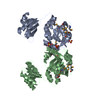
| ||||||||
|---|---|---|---|---|---|---|---|---|---|
| 1 | 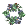
| ||||||||
| Unit cell |
|
-Components
| #1: Protein | Mass: 27149.689 Da / Num. of mol.: 2 Source method: isolated from a genetically manipulated source Source: (gene. exp.) Haemophilus influenzae (bacteria) / Plasmid: pET17b / Production host: #2: Chemical | ChemComp-SO4 /   Mass: 96.063 Da / Num. of mol.: 4 / Source method: obtained synthetically / Formula: SO4 Mass: 96.063 Da / Num. of mol.: 4 / Source method: obtained synthetically / Formula: SO4 |
|---|
-Experimental details
-Experiment
| Experiment | Method: X-RAY DIFFRACTION / Number of used crystals: 1 |
|---|
- Sample preparation
| Crystal | Density Matthews: 2.91 Å3/Da / Density % sol: 57.44 % | ||||||||||||||||||||||||||||||
|---|---|---|---|---|---|---|---|---|---|---|---|---|---|---|---|---|---|---|---|---|---|---|---|---|---|---|---|---|---|---|---|
| Crystal grow | Temperature: 298 K / Method: vapor diffusion, hanging drop / pH: 8 Details: Ammonium sulfate, sodium acetate, tris, pH 8.0, VAPOR DIFFUSION, HANGING DROP, temperature 298K | ||||||||||||||||||||||||||||||
| Crystal grow | *PLUS Method: vapor diffusion | ||||||||||||||||||||||||||||||
| Components of the solutions | *PLUS
|
-Data collection
| Diffraction | Mean temperature: 180 K |
|---|---|
| Diffraction source | Source: SYNCHROTRON / Site: Photon Factory  / Beamline: BL-18B / Wavelength: 0.9795 Å / Beamline: BL-18B / Wavelength: 0.9795 Å |
| Detector | Type: ADSC QUANTUM 4 / Detector: CCD / Date: Nov 22, 2001 / Details: mirrors |
| Radiation | Monochromator: mirrors / Protocol: MAD / Monochromatic (M) / Laue (L): M / Scattering type: x-ray |
| Radiation wavelength | Wavelength: 0.9795 Å / Relative weight: 1 |
| Reflection | Resolution: 2.8→99 Å / Num. all: 6059 / Num. obs: 15821 / % possible obs: 98.5 % / Observed criterion σ(F): 0 / Observed criterion σ(I): 0 |
| Reflection shell | Resolution: 2.8→99 Å / % possible all: 98.5 |
| Reflection | *PLUS Num. obs: 15906 / Num. measured all: 407938 / Rmerge(I) obs: 0.085 |
| Reflection shell | *PLUS Highest resolution: 2.8 Å / Lowest resolution: 2.95 Å / % possible obs: 100 % / Rmerge(I) obs: 0.264 / Mean I/σ(I) obs: 2.8 |
- Processing
| Software |
| ||||||||||||||||||||||||
|---|---|---|---|---|---|---|---|---|---|---|---|---|---|---|---|---|---|---|---|---|---|---|---|---|---|
| Refinement | Method to determine structure: MAD / Resolution: 2.8→99 Å / σ(F): 0 / σ(I): 0 / Stereochemistry target values: Engh & Huber
| ||||||||||||||||||||||||
| Refinement step | Cycle: LAST / Resolution: 2.8→99 Å
| ||||||||||||||||||||||||
| Refinement | *PLUS Lowest resolution: 99 Å / % reflection Rfree: 5 % | ||||||||||||||||||||||||
| Solvent computation | *PLUS | ||||||||||||||||||||||||
| Displacement parameters | *PLUS | ||||||||||||||||||||||||
| Refine LS restraints | *PLUS
|
