 Movie
Movie Controller

[English] 日本語
 Yorodumi
- PDB-1nko: Energetic and structural basis of sialylated oligosaccharide reco... -
+ Open data

- Basic information
| Entry | Database: PDB / ID: 1nko | ||||||
|---|---|---|---|---|---|---|---|
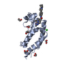
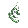
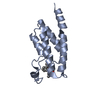
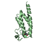
| Title | Energetic and structural basis of sialylated oligosaccharide recognition by the natural killer cell inhibitory receptor p75/AIRM1 or Siglec-7 | ||||||
 Components Components | Sialic acid binding Ig-like lectin 7 | ||||||
 Keywords Keywords | IMMUNE SYSTEM / Immunoglobulin / Siglec7 | ||||||
| Function / homology |  Function and homology information Function and homology informationsialic acid binding / Immunoregulatory interactions between a Lymphoid and a non-Lymphoid cell / signaling receptor activity / carbohydrate binding / cell adhesion / plasma membrane Similarity search - Function | ||||||
| Biological species |  Homo sapiens (human) Homo sapiens (human) | ||||||
| Method |  X-RAY DIFFRACTION / SYNCHROTRON / MOLECULAR REPLACEMENT / Resolution: 1.45 Å X-RAY DIFFRACTION / SYNCHROTRON / MOLECULAR REPLACEMENT / Resolution: 1.45 Å | ||||||
 Authors Authors | Dimasi, N. / Attril, H. / van Aalten, D.M.F. / Moretta, L. / Biassoni, R. / Mariuzza, R.A. | ||||||
 Citation Citation | Journal: Acta Crystallogr.,Sect.D / Year: 2004 Title: Structure of the saccharide-binding domain of the human natural killer cell inhibitory receptor p75/AIRM1. Authors: Dimasi, N. / Moretta, A. / Moretta, L. / Biassoni, R. / Mariuzza, R.A. | ||||||
| History |
|
- Structure visualization
| Structure viewer | Molecule: MolmilJmol/JSmol |
|---|
- Downloads & links
-Download
| PDBx/mmCIF format | 1nko.cif.gz | 37.5 KB | Display | PDBx/mmCIF format |
|---|---|---|---|---|
| PDB format | pdb1nko.ent.gz | 24.9 KB | Display | PDB format |
| PDBx/mmJSON format | 1nko.json.gz | Tree view | PDBx/mmJSON format | |
| Others |  Other downloads Other downloads |
-Validation report
| Summary document | 1nko_validation.pdf.gz | 431.4 KB | Display | wwPDB validaton report |
|---|---|---|---|---|
| Full document | 1nko_full_validation.pdf.gz | 435 KB | Display | |
| Data in XML | 1nko_validation.xml.gz | 7 KB | Display | |
| Data in CIF | 1nko_validation.cif.gz | 8.3 KB | Display | |
| Arichive directory | https://data.pdbj.org/pub/pdb/validation_reports/nk/1nkoftp://data.pdbj.org/pub/pdb/validation_reports/nk/1nko | HTTPS FTP |
-Related structure data
| Related structure data |  1qfoS S: Starting model for refinement |
|---|---|
| Similar structure data |


-Links
 PDBj
PDBj
- Assembly
| Deposited unit | 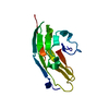
| ||||||||
|---|---|---|---|---|---|---|---|---|---|
| 1 |
| ||||||||
| Unit cell |
|
-Components
| #1: Protein | Mass: 15262.995 Da / Num. of mol.: 1 / Fragment: N-terminal IgV-like domain residues 19-150 / Mutation: Q55D Source method: isolated from a genetically manipulated source Source: (gene. exp.) Homo sapiens (human) / Gene: SIGLEC7 OR AIRM1 / Plasmid: p75-1D / Production host:  |
|---|
-Experimental details
-Experiment
| Experiment | Method: X-RAY DIFFRACTION / Number of used crystals: 1 |
|---|
- Sample preparation
| Crystal | Density Matthews: 1.94 Å3/Da / Density % sol: 36.43 % | ||||||||||||||||||||||||||||||||||||||||||
|---|---|---|---|---|---|---|---|---|---|---|---|---|---|---|---|---|---|---|---|---|---|---|---|---|---|---|---|---|---|---|---|---|---|---|---|---|---|---|---|---|---|---|---|
| Crystal grow | Temperature: 298 K / Method: vapor diffusion / pH: 4.2 Details: PEG 4000 20%, NaCl 0.2 M, 0.1 M phosphate-citrate, pH 4.2, VAPOR DIFFUSION, temperature 298K | ||||||||||||||||||||||||||||||||||||||||||
| Crystal grow | *PLUS Temperature: 277 K / pH: 5.5 / Method: vapor diffusion, hanging drop / Details: Dimasi, N., (2003) Acta Crystallogr., D59, 1856. | ||||||||||||||||||||||||||||||||||||||||||
| Components of the solutions | *PLUS
|
-Data collection
| Diffraction | Mean temperature: 100 K |
|---|---|
| Diffraction source | Source: SYNCHROTRON / Site: CHESS  / Beamline: F1 / Wavelength: 0.9 Å / Beamline: F1 / Wavelength: 0.9 Å |
| Detector | Type: ADSC QUANTUM 4 / Detector: CCD / Date: May 20, 2000 |
| Radiation | Monochromator: GRAPHITE / Protocol: SINGLE WAVELENGTH / Monochromatic (M) / Laue (L): M / Scattering type: x-ray |
| Radiation wavelength | Wavelength: 0.9 Å / Relative weight: 1 |
| Reflection | Resolution: 1.45→15 Å / Num. obs: 19800 / % possible obs: 75 % / Observed criterion σ(F): 2 / Observed criterion σ(I): 2 / Biso Wilson estimate: 19.8 Å2 |
| Reflection shell | Resolution: 1.45→15 Å / % possible all: 95.3 |
| Reflection | *PLUS Lowest resolution: 15 Å / Num. obs: 20819 / % possible obs: 89.3 % / Num. measured all: 238401 / Rmerge(I) obs: 0.06 |
| Reflection shell | *PLUS Lowest resolution: 1.51 Å / % possible obs: 68.6 % / Rmerge(I) obs: 0.388 / Mean I/σ(I) obs: 2.5 |
- Processing
| Software |
| ||||||||||||||||||||||||||||||||||||
|---|---|---|---|---|---|---|---|---|---|---|---|---|---|---|---|---|---|---|---|---|---|---|---|---|---|---|---|---|---|---|---|---|---|---|---|---|---|
| Refinement | Method to determine structure: MOLECULAR REPLACEMENT Starting model: 1QFO Resolution: 1.45→14.93 Å / Rfactor Rfree error: 0.01 / Isotropic thermal model: RESTRAINED / Cross valid method: THROUGHOUT / σ(F): 2 / Stereochemistry target values: Engh & Huber
| ||||||||||||||||||||||||||||||||||||
| Solvent computation | Solvent model: FLAT MODEL / Bsol: 54.3265 Å2 / ksol: 0.44952 e/Å3 | ||||||||||||||||||||||||||||||||||||
| Displacement parameters | Biso mean: 21.5 Å2
| ||||||||||||||||||||||||||||||||||||
| Refine analyze | Luzzati coordinate error free: 0.26 Å / Luzzati sigma a free: 0.21 Å | ||||||||||||||||||||||||||||||||||||
| Refinement step | Cycle: LAST / Resolution: 1.45→14.93 Å
| ||||||||||||||||||||||||||||||||||||
| Refine LS restraints |
| ||||||||||||||||||||||||||||||||||||
| LS refinement shell | Resolution: 1.45→1.54 Å / Rfactor Rfree error: 0.036 / Total num. of bins used: 6
| ||||||||||||||||||||||||||||||||||||
| Xplor file |
| ||||||||||||||||||||||||||||||||||||
| Refinement | *PLUS Lowest resolution: 15 Å / % reflection Rfree: 4 % / Rfactor Rfree: 0.218 / Rfactor Rwork: 0.171 | ||||||||||||||||||||||||||||||||||||
| Solvent computation | *PLUS | ||||||||||||||||||||||||||||||||||||
| Displacement parameters | *PLUS | ||||||||||||||||||||||||||||||||||||
| Refine LS restraints | *PLUS
|
