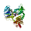
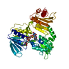
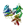
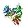
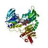
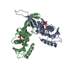
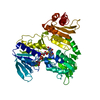
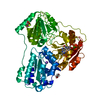
登録情報 データベース : PDB / ID : 1nhqタイトル CRYSTALLOGRAPHIC ANALYSES OF NADH PEROXIDASE CYS42ALA AND CYS42SER MUTANTS: ACTIVE SITE STRUCTURE, MECHANISTIC IMPLICATIONS, AND AN UNUSUAL ENVIRONMENT OF ARG303 NADH PEROXIDASE キーワード 機能・相同性 分子機能 ドメイン・相同性 構成要素
/ / / / / / / / / / / / / / / / / / / / / / / / 生物種 Enterococcus faecalis (乳酸球菌)手法 / 解像度 : 2 Å データ登録者 Mande, S.S. / Claiborne, A. / Hol, W.G.J. 履歴 登録 1994年12月9日 処理サイト 改定 1.0 1995年2月14日 Provider / タイプ 改定 1.1 2008年3月24日 Group 改定 1.2 2011年7月13日 Group / Version format compliance改定 1.3 2012年3月28日 Group 改定 1.4 2024年2月14日 Group Data collection / Database references ... Data collection / Database references / Derived calculations / Other カテゴリ chem_comp_atom / chem_comp_bond ... chem_comp_atom / chem_comp_bond / database_2 / pdbx_database_status / struct_ref_seq_dif / struct_site Item _database_2.pdbx_DOI / _database_2.pdbx_database_accession ... _database_2.pdbx_DOI / _database_2.pdbx_database_accession / _pdbx_database_status.process_site / _struct_ref_seq_dif.details / _struct_site.pdbx_auth_asym_id / _struct_site.pdbx_auth_comp_id / _struct_site.pdbx_auth_seq_id
すべて表示 表示を減らす
 ムービー
ムービー コントローラー


 データを開く
 基本情報
基本情報 要素
要素 キーワード
キーワード 機能・相同性情報
 Enterococcus faecalis (乳酸球菌)
 X線回折 / 解像度: 2 Å
  データ登録者
 引用
引用 構造の表示
構造の表示 ダウンロードとリンク
 その他のダウンロード


 PDBj
PDBj


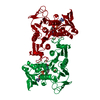
 集合体
集合体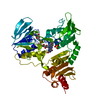

 分子量: 96.063 Da / 分子数: 1 / 由来タイプ: 合成 / 式: SO4
 分子量: 785.550 Da / 分子数: 1 / 由来タイプ: 合成 / 式: C27H33N9O15P2 / コメント: FAD*YM
 分子量: 18.015 Da / 分子数: 309 / 由来タイプ: 天然 / 式: H2O
 試料調製
試料調製 解析
解析