 Movie
Movie Controller

[English] 日本語
 Yorodumi
- PDB-1nb3: Crystal structure of stefin A in complex with cathepsin H: N-term... -
+ Open data

- Basic information
| Entry | Database: PDB / ID: 1nb3 | |||||||||
|---|---|---|---|---|---|---|---|---|---|---|
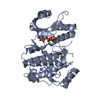
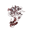
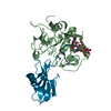
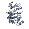
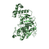
| Title | Crystal structure of stefin A in complex with cathepsin H: N-terminal residues of inhibitors can adapt to the active sites of endo-and exopeptidases | |||||||||
 Components Components |
| |||||||||
 Keywords Keywords | HYDROLASE / CYSTEINE PROTEINASE / AMINOPEPTIDASE / CYSTATIN / ENZYME-INHIBITOR COMPLEX | |||||||||
| Function / homology |  Function and homology information Function and homology informationcathepsin H / neuropeptide catabolic process / HLA-A specific activating MHC class I receptor activity / dichotomous subdivision of terminal units involved in lung branching / positive regulation of peptidase activity / Surfactant metabolism / alveolar lamellar body / negative regulation of peptidase activity / immune response-regulating signaling pathway / peptidase inhibitor complex ...cathepsin H / neuropeptide catabolic process / HLA-A specific activating MHC class I receptor activity / dichotomous subdivision of terminal units involved in lung branching / positive regulation of peptidase activity / Surfactant metabolism / alveolar lamellar body / negative regulation of peptidase activity / immune response-regulating signaling pathway / peptidase inhibitor complex / membrane protein proteolysis / Formation of the cornified envelope / bradykinin catabolic process / peptide cross-linking / cornified envelope / Neutrophil degranulation / metanephros development / surfactant homeostasis / zymogen activation / MHC class II antigen presentation / cysteine-type endopeptidase inhibitor activity / positive regulation of epithelial cell migration / thyroid hormone binding / cysteine-type endopeptidase activator activity involved in apoptotic process / aminopeptidase activity / response to retinoic acid / cysteine-type peptidase activity / keratinocyte differentiation / ERK1 and ERK2 cascade / positive regulation of apoptotic signaling pathway / proteolysis involved in protein catabolic process / negative regulation of proteolysis / cell-cell adhesion / protein destabilization / T cell mediated cytotoxicity / positive regulation of angiogenesis / endopeptidase activity / protease binding / lysosome / positive regulation of cell migration / immune response / serine-type endopeptidase activity / cysteine-type endopeptidase activity / positive regulation of cell population proliferation / positive regulation of gene expression / negative regulation of apoptotic process / proteolysis / extracellular space / nucleoplasm / cytoplasm / cytosol Similarity search - Function | |||||||||
| Biological species |  Homo sapiens (human) Homo sapiens (human) | |||||||||
| Method |  X-RAY DIFFRACTION / MOLECULAR REPLACEMENT / Resolution: 2.8 Å X-RAY DIFFRACTION / MOLECULAR REPLACEMENT / Resolution: 2.8 Å | |||||||||
 Authors Authors | Jenko, S. / Dolenc, I. / Guncar, G. / Dobersek, A. / Podobnik, M. / Turk, D. | |||||||||
 Citation Citation | Journal: J.Mol.Biol. / Year: 2003 Title: Crystal structure of stefin A in complex with cathepsin H: N-terminal residues of inhibitors can adapt to the active sites of endo- and exopeptidases Authors: Jenko, S. / Dolenc, I. / Guncar, G. / Dobersek, A. / Podobnik, M. / Turk, D. | |||||||||
| History |
|
- Structure visualization
| Structure viewer | Molecule: MolmilJmol/JSmol |
|---|
- Downloads & links
-Download
| PDBx/mmCIF format | 1nb3.cif.gz | 276.1 KB | Display | PDBx/mmCIF format |
|---|---|---|---|---|
| PDB format | pdb1nb3.ent.gz | 222.4 KB | Display | PDB format |
| PDBx/mmJSON format | 1nb3.json.gz | Tree view | PDBx/mmJSON format | |
| Others |  Other downloads Other downloads |
-Validation report
| Summary document | 1nb3_validation.pdf.gz | 1.6 MB | Display | wwPDB validaton report |
|---|---|---|---|---|
| Full document | 1nb3_full_validation.pdf.gz | 1.7 MB | Display | |
| Data in XML | 1nb3_validation.xml.gz | 61.8 KB | Display | |
| Data in CIF | 1nb3_validation.cif.gz | 87.1 KB | Display | |
| Arichive directory | https://data.pdbj.org/pub/pdb/validation_reports/nb/1nb3ftp://data.pdbj.org/pub/pdb/validation_reports/nb/1nb3 | HTTPS FTP |
-Related structure data
| Related structure data |  1nb5C 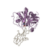 1stfS  8pchS C: citing same article ( S: Starting model for refinement |
|---|---|
| Similar structure data |


-Links
 PDBj
PDBj

- Assembly
| Deposited unit | 
| ||||||||
|---|---|---|---|---|---|---|---|---|---|
| 1 | 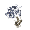
| ||||||||
| 2 | 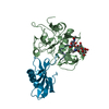
| ||||||||
| 3 | 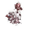
| ||||||||
| 4 | 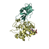
| ||||||||
| Unit cell |
|
-Components
| #1: Protein | Mass: 24328.521 Da / Num. of mol.: 4 / Source method: isolated from a natural source / Details: protein was isolated from spleen / Source: (natural) #2: Protein/peptide | Mass: 848.878 Da / Num. of mol.: 4 / Source method: isolated from a natural source / Details: protein was isolated from spleen / Source: (natural) #3: Protein | Mass: 11020.464 Da / Num. of mol.: 4 Source method: isolated from a genetically manipulated source Source: (gene. exp.) Homo sapiens (human) / Gene: CSTA OR STF1 / Plasmid: pET3a / Production host:  #4: Polysaccharide | beta-D-mannopyranose-(1-4)-2-acetamido-2-deoxy-beta-D-glucopyranose-(1-4)-2-acetamido-2-deoxy-beta- ...beta-D-mannopyranose-(1-4)-2-acetamido-2-deoxy-beta-D-glucopyranose-(1-4)-2-acetamido-2-deoxy-beta-D-glucopyranose Source method: isolated from a genetically manipulated source #5: Water | ChemComp-HOH / |  Mass: 18.015 Da / Num. of mol.: 543 / Source method: isolated from a natural source / Formula: H2O Mass: 18.015 Da / Num. of mol.: 543 / Source method: isolated from a natural source / Formula: H2O |
|---|
-Experimental details
-Experiment
| Experiment | Method: X-RAY DIFFRACTION / Number of used crystals: 1 |
|---|
- Sample preparation
| Crystal | Density Matthews: 2.36 Å3/Da / Density % sol: 47.39 % | ||||||||||||||||||||||||||||||||||||||||||
|---|---|---|---|---|---|---|---|---|---|---|---|---|---|---|---|---|---|---|---|---|---|---|---|---|---|---|---|---|---|---|---|---|---|---|---|---|---|---|---|---|---|---|---|
| Crystal grow | *PLUS Temperature: 22 ℃ / pH: 4.2 / Method: vapor diffusion | ||||||||||||||||||||||||||||||||||||||||||
| Components of the solutions | *PLUS
|
-Data collection
| Diffraction | Mean temperature: 90 K |
|---|---|
| Diffraction source | Source: ROTATING ANODE / Type: RIGAKU RU200 / Wavelength: 1.5418 Å |
| Detector | Type: MARRESEARCH / Detector: IMAGE PLATE / Date: Jan 8, 2001 / Details: mirrors |
| Radiation | Monochromator: YALE MIRRORS / Protocol: SINGLE WAVELENGTH / Monochromatic (M) / Laue (L): M / Scattering type: x-ray |
| Radiation wavelength | Wavelength: 1.5418 Å / Relative weight: 1 |
| Reflection | Resolution: 2.8→99 Å / Num. obs: 34127 / % possible obs: 97.1 % / Observed criterion σ(F): 0 / Observed criterion σ(I): -3 / Redundancy: 8.9 % / Rsym value: 0.161 |
| Reflection shell | Resolution: 2.8→2.9 Å / Redundancy: 2.3 % / Rmerge(I) obs: 0.161 / Mean I/σ(I) obs: 4.7 / Num. unique all: 3405 / Rsym value: 0.583 / % possible all: 97.4 |
| Reflection | *PLUS Lowest resolution: 99 Å / Num. obs: 35154 / % possible obs: 97.4 % / Num. measured all: 313477 / Rmerge(I) obs: 0.161 |
| Reflection shell | *PLUS % possible obs: 97.1 % / Rmerge(I) obs: 0.583 |
- Processing
| Software |
| ||||||||||||||||||||
|---|---|---|---|---|---|---|---|---|---|---|---|---|---|---|---|---|---|---|---|---|---|
| Refinement | Method to determine structure: MOLECULAR REPLACEMENT Starting model: PDB ENTRY 1STF, 8PCH Resolution: 2.8→10 Å / Cross valid method: R-FREE, KICKED OMIT MAP / σ(F): 0
| ||||||||||||||||||||
| Refinement step | Cycle: LAST / Resolution: 2.8→10 Å
| ||||||||||||||||||||
| Refine LS restraints |
| ||||||||||||||||||||
| LS refinement shell | Highest resolution: 2.8 Å
| ||||||||||||||||||||
| Refinement | *PLUS Lowest resolution: 10 Å / % reflection Rfree: 10 % / Rfactor Rwork: 0.228 | ||||||||||||||||||||
| Solvent computation | *PLUS | ||||||||||||||||||||
| Displacement parameters | *PLUS | ||||||||||||||||||||
| LS refinement shell | *PLUS Highest resolution: 2.8 Å |
