- PDB-1n68: Copper bound to the Multicopper Oxidase CueO -
+
Open data
ID or keywords:
Loading...
-
Basic information
Entry
Database: PDB / ID: 1n68
Title
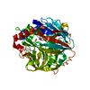
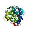
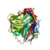
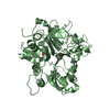
Copper bound to the Multicopper Oxidase CueO
Components
Blue copper oxidase cueO
Keywords
OXIDOREDUCTASE / copper / multicopper oxidase
Function / homology
Function and homology information
cuproxidase / oxidoreductase activity, acting on metal ions, oxygen as acceptor / oxidoreductase activity, acting on metal ions / detoxification of copper ion / oxidoreductase activity, acting on diphenols and related substances as donors, oxygen as acceptor / response to copper ion / ferroxidase activity / outer membrane-bounded periplasmic space / periplasmic space / copper ion binding Similarity search - Function
Resolution: 1.7→26.3 Å / Isotropic thermal model: Isotropic / Cross valid method: THROUGHOUT / σ(F): 0 / σ(I): 0 / Stereochemistry target values: Engh & Huber Details: Missing from the model are residues 1-29 at the N-terminus (1-28 are a presumably cleaved signal peptide), and residues 380-402 which are not visible in the electron density map and ...Details: Missing from the model are residues 1-29 at the N-terminus (1-28 are a presumably cleaved signal peptide), and residues 380-402 which are not visible in the electron density map and presumably disordered. Metal-ligand bond distances and angles were not restrained.
Rfactor
Num. reflection
% reflection
Selection details
Rfree
0.222
2604
-
RANDOM
Rwork
0.165
-
-
-
all
0.171
51492
-
-
obs
0.171
51492
99.7 %
-
Displacement parameters
Biso mean: 21.4 Å2
Refinement step
Cycle: LAST / Resolution: 1.7→26.3 Å
Protein
Nucleic acid
Ligand
Solvent
Total
Num. atoms
3568
0
7
452
4027
Refine LS restraints
Refine-ID
Type
Dev ideal
X-RAY DIFFRACTION
s_bond_d
0.007
X-RAY DIFFRACTION
s_angle_d
0.024
X-RAY DIFFRACTION
s_from_restr_planes
0.0287
+
About Yorodumi
-
News
-
Feb 9, 2022. New format data for meta-information of EMDB entries
New format data for meta-information of EMDB entries
Version 3 of the EMDB header file is now the official format.
The previous official version 1.9 will be removed from the archive.
In the structure databanks used in Yorodumi, some data are registered as the other names, "COVID-19 virus" and "2019-nCoV". Here are the details of the virus and the list of structure data.
Jan 31, 2019. EMDB accession codes are about to change! (news from PDBe EMDB page)
EMDB accession codes are about to change! (news from PDBe EMDB page)
The allocation of 4 digits for EMDB accession codes will soon come to an end. Whilst these codes will remain in use, new EMDB accession codes will include an additional digit and will expand incrementally as the available range of codes is exhausted. The current 4-digit format prefixed with “EMD-” (i.e. EMD-XXXX) will advance to a 5-digit format (i.e. EMD-XXXXX), and so on. It is currently estimated that the 4-digit codes will be depleted around Spring 2019, at which point the 5-digit format will come into force.
The EM Navigator/Yorodumi systems omit the EMD- prefix.
Related info.:Q: What is EMD? / ID/Accession-code notation in Yorodumi/EM Navigator
Yorodumi is a browser for structure data from EMDB, PDB, SASBDB, etc.
This page is also the successor to EM Navigator detail page, and also detail information page/front-end page for Omokage search.
The word "yorodu" (or yorozu) is an old Japanese word meaning "ten thousand". "mi" (miru) is to see.
Related info.:EMDB / PDB / SASBDB / Comparison of 3 databanks / Yorodumi Search / Aug 31, 2016. New EM Navigator & Yorodumi / Yorodumi Papers / Jmol/JSmol / Function and homology information / Changes in new EM Navigator and Yorodumi
 Movie
Movie Controller


 Open data
 Basic information
 Components
 Keywords
 Function and homology information
 X-RAY DIFFRACTION /
  Authors
 Citation
 Structure visualization
 Downloads & links
 Other downloads


 PDBj
PDBj

 Assembly

 Mass: 63.546 Da / Num. of mol.: 4 / Source method: obtained synthetically / Formula: Cu
 Mass: 162.545 Da / Num. of mol.: 1 / Source method: obtained synthetically / Formula: ClCu2
 Mass: 18.015 Da / Num. of mol.: 452 / Source method: isolated from a natural source / Formula: H2O
 Sample preparation
 / Beamline: BL9-2 / Wavelength: 1.378 Å
 Processing