 Movie
Movie Controller

[English] 日本語
 Yorodumi
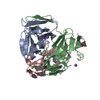
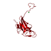
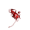
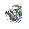
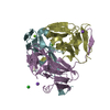
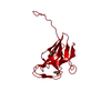
- PDB-1mq7: CRYSTAL STRUCTURE OF DUTPASE FROM MYCOBACTERIUM TUBERCULOSIS (RV2697C) -
+ Open data

- Basic information
| Entry | Database: PDB / ID: 1mq7 | ||||||
|---|---|---|---|---|---|---|---|
| Title | CRYSTAL STRUCTURE OF DUTPASE FROM MYCOBACTERIUM TUBERCULOSIS (RV2697C) | ||||||
 Components Components | DEOXYURIDINE 5'-TRIPHOSPHATE NUCLEOTIDOHYDROLASE | ||||||
 Keywords Keywords | HYDROLASE / jelly-roll / Structural Genomics / PSI / Protein Structure Initiative / TB Structural Genomics Consortium / TBSGC | ||||||
| Function / homology |  Function and homology information Function and homology informationdUTP metabolic process / dUTP catabolic process / dUMP biosynthetic process / dUTP diphosphatase / dUTP diphosphatase activity / magnesium ion binding Similarity search - Function | ||||||
| Biological species |   Mycobacterium tuberculosis (bacteria) Mycobacterium tuberculosis (bacteria) | ||||||
| Method |  X-RAY DIFFRACTION / SYNCHROTRON / MOLECULAR REPLACEMENT / Resolution: 1.95 Å X-RAY DIFFRACTION / SYNCHROTRON / MOLECULAR REPLACEMENT / Resolution: 1.95 Å | ||||||
 Authors Authors | Sawaya, M.R. / Chan, S. / Segelke, B.W. / Lekin, T. / Heike, K. / Cho, U.S. / Naranjo, C. / Perry, L.J. / Yeates, T.O. / Eisenberg, D. / TB Structural Genomics Consortium (TBSGC) | ||||||
 Citation Citation | Journal: J.Mol.Biol. / Year: 2004 Title: Crystal structure of the Mycobacterium tuberculosis dUTPase: insights into the catalytic mechanism. Authors: Chan, S. / Segelke, B. / Lekin, T. / Krupka, H. / Cho, U.S. / Kim, M.-Y. / So, M. / Kim, C.-Y. / Naranjo, C.M. / Rogers, Y.C. / Park, M.S. / Waldo, G.S. / Pashkov, I. / Cascio, D. / Perry, J.L. / Sawaya, M.R. #1: Journal: STRUCTURE / Year: 1996Title: HUMAN DUTP PYROPHOSPHATASE: URACIL RECOGNITION BY A BETA HAIRPIN AND ACTIVE SITES FORMED BY THREE SEPARATE SUBUNITS Authors: Mol, C.D. / Harris, J.M. / Mclntosh, E.M. / Tainer, J.A. | ||||||
| History |
|
- Structure visualization
| Structure viewer | Molecule: MolmilJmol/JSmol |
|---|
- Downloads & links
-Download
| PDBx/mmCIF format | 1mq7.cif.gz | 41 KB | Display | PDBx/mmCIF format |
|---|---|---|---|---|
| PDB format | pdb1mq7.ent.gz | 28.2 KB | Display | PDB format |
| PDBx/mmJSON format | 1mq7.json.gz | Tree view | PDBx/mmJSON format | |
| Others |  Other downloads Other downloads |
-Validation report
| Arichive directory | https://data.pdbj.org/pub/pdb/validation_reports/mq/1mq7ftp://data.pdbj.org/pub/pdb/validation_reports/mq/1mq7 | HTTPS FTP |
|---|
-Related structure data
| Related structure data | 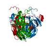 1sixC 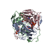 1sjnC 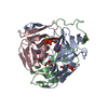 1slhC  1sm8C 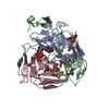 1smcC 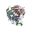 1snfC 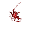 1dudS S: Starting model for refinement C: citing same article ( |
|---|---|
| Similar structure data | |
| Other databases |


-Links
 PDBj
PDBj
- Assembly
| Deposited unit | 
| |||||||||||||||||||||
|---|---|---|---|---|---|---|---|---|---|---|---|---|---|---|---|---|---|---|---|---|---|---|
| 1 | 
| |||||||||||||||||||||
| Unit cell |
| |||||||||||||||||||||
| Components on special symmetry positions |
| |||||||||||||||||||||
| Details | The biological assembly is a trimer that is generated by the following crystallographic symmetry operators -y+1, -z+1, x and z, -x+1, -y+1 |
-Components
| #1: Protein | Mass: 15820.938 Da / Num. of mol.: 1 Source method: isolated from a genetically manipulated source Source: (gene. exp.) Mycobacterium tuberculosis (bacteria) / Gene: Rv2697c / Production host: References: UniProt: P0A552, UniProt: P9WNS5*PLUS, dUTP diphosphatase | ||
|---|---|---|---|
| #2: Chemical |   Mass: 122.143 Da / Num. of mol.: 2 / Source method: obtained synthetically / Formula: C4H12NO3 / Comment: pH buffer*YM Mass: 122.143 Da / Num. of mol.: 2 / Source method: obtained synthetically / Formula: C4H12NO3 / Comment: pH buffer*YM#3: Water | ChemComp-HOH / |  Mass: 18.015 Da / Num. of mol.: 135 / Source method: isolated from a natural source / Formula: H2O Mass: 18.015 Da / Num. of mol.: 135 / Source method: isolated from a natural source / Formula: H2O |
-Experimental details
-Experiment
| Experiment | Method: X-RAY DIFFRACTION / Number of used crystals: 1 |
|---|
- Sample preparation
| Crystal | Density Matthews: 2.54 Å3/Da / Density % sol: 51.58 % | ||||||||||||||||||||||||||||||
|---|---|---|---|---|---|---|---|---|---|---|---|---|---|---|---|---|---|---|---|---|---|---|---|---|---|---|---|---|---|---|---|
| Crystal grow | Temperature: 298 K / Method: vapor diffusion, hanging drop / pH: 8.5 Details: PEG4000, EDTA, Tris, pH 8.5, VAPOR DIFFUSION, HANGING DROP, temperature 298K | ||||||||||||||||||||||||||||||
| Crystal grow | *PLUS Method: vapor diffusion, sitting drop | ||||||||||||||||||||||||||||||
| Components of the solutions | *PLUS
|
-Data collection
| Diffraction | Mean temperature: 100 K |
|---|---|
| Diffraction source | Source: SYNCHROTRON / Site: ALS  / Beamline: 5.0.1 / Wavelength: 1 Å / Beamline: 5.0.1 / Wavelength: 1 Å |
| Detector | Type: ADSC QUANTUM 4 / Detector: CCD / Date: Jun 23, 2002 |
| Radiation | Monochromator: Si (220) / Protocol: SINGLE WAVELENGTH / Monochromatic (M) / Laue (L): M / Scattering type: x-ray |
| Radiation wavelength | Wavelength: 1 Å / Relative weight: 1 |
| Reflection | Resolution: 1.95→31.25 Å / Num. all: 11317 / Num. obs: 11317 / % possible obs: 97.1 % / Observed criterion σ(F): 0 / Observed criterion σ(I): 0 / Redundancy: 18 % / Biso Wilson estimate: 17.3 Å2 / Rsym value: 0.078 / Net I/σ(I): 62.7 |
| Reflection shell | Resolution: 1.95→2.02 Å / Num. unique all: 1172 / Rsym value: 0.393 / % possible all: 100 |
| Reflection | *PLUS Num. obs: 11592 / Num. measured all: 208809 / Rmerge(I) obs: 0.078 |
| Reflection shell | *PLUS % possible obs: 100 % / Rmerge(I) obs: 0.393 / Mean I/σ(I) obs: 13.5 |
- Processing
| Software |
| |||||||||||||||||||||||||
|---|---|---|---|---|---|---|---|---|---|---|---|---|---|---|---|---|---|---|---|---|---|---|---|---|---|---|
| Refinement | Method to determine structure: MOLECULAR REPLACEMENT Starting model: PDB ENTRY 1DUD Resolution: 1.95→31.25 Å / Rfactor Rfree error: 0.007 / Isotropic thermal model: RESTRAINED / Cross valid method: THROUGHOUT / σ(F): 0 / Stereochemistry target values: Engh & Huber
| |||||||||||||||||||||||||
| Solvent computation | Solvent model: FLAT MODEL / Bsol: 48.1896 Å2 / ksol: 0.366749 e/Å3 | |||||||||||||||||||||||||
| Displacement parameters | Biso mean: 31.8 Å2
| |||||||||||||||||||||||||
| Refine analyze | Luzzati coordinate error free: 0.26 Å / Luzzati sigma a free: 0.14 Å | |||||||||||||||||||||||||
| Refinement step | Cycle: LAST / Resolution: 1.95→31.25 Å
| |||||||||||||||||||||||||
| Refine LS restraints |
| |||||||||||||||||||||||||
| LS refinement shell | Resolution: 1.95→2.07 Å / Rfactor Rfree error: 0.018 / Total num. of bins used: 6
| |||||||||||||||||||||||||
| Xplor file |
| |||||||||||||||||||||||||
| Refinement | *PLUS % reflection Rfree: 5 % | |||||||||||||||||||||||||
| Solvent computation | *PLUS | |||||||||||||||||||||||||
| Displacement parameters | *PLUS | |||||||||||||||||||||||||
| Refine LS restraints | *PLUS
|
